Review
. 2015 Sep 19;370(1677):20140206.
doi: 10.1098/rstb.2014.0206.

Playing the electric light orchestra--how electrical stimulation of visual cortex elucidates the neural basis of perception

Affiliations
Review

Playing the electric light orchestra--how electrical stimulation of visual cortex elucidates the neural basis of perception

Nela Cicmil et al. Philos Trans R Soc Lond B Biol Sci. .

Abstract

Vision research has the potential to reveal fundamental mechanisms underlying sensory experience. Causal experimental approaches, such as electrical microstimulation, provide a unique opportunity to test the direct contributions of visual cortical neurons to perception and behaviour. But in spite of their importance, causal methods constitute a minority of the experiments used to investigate the visual cortex to date. We reconsider the function and organization of visual cortex according to results obtained from stimulation techniques, with a special emphasis on electrical stimulation of small groups of cells in awake subjects who can report their visual experience. We compare findings from humans and monkeys, striate and extrastriate cortex, and superficial versus deep cortical layers, and identify a number of revealing gaps in the 'causal map' of visual cortex. Integrating results from different methods and species, we provide a critical overview of the ways in which causal approaches have been used to further our understanding of circuitry, plasticity and information integration in visual cortex. Electrical stimulation not only elucidates the contributions of different visual areas to perception, but also contributes to our understanding of neuronal mechanisms underlying memory, attention and decision-making.

Keywords: decision-making; electrical stimulation; optogenetics; perception; primate; visual cortex.


Figures

Figure 1.
Figure 1.
Overview of sites where causal stimulation experiments have been performed in the visual cortex (and selected connected areas) of humans and monkeys (see also tables 1 and 2). Sites are shown on schematic human and macaque brains, and indicate the visual cortical areas involved (not exact electrode positions). (a) Visual cortical sites of electrical stimulation in human patients where either a simple phosphene percept was evoked with a cortical surface electrode (red triangle) or with an intracortical microelectrode (red cross), or where a complex form percept was evoked (purple triangle—surface electrodes only). Most sites where larger currents evoke reportable percepts are around primary visual cortex (V1) and the fusiform face area (FFA). (b) Visual cortical sites for which macaque monkeys have detected intracortical electrical microstimulation either without (red cross) or with extensive training to specifically detect electrical microstimulation (blue cross). For extrastriate visual cortex, specific detection training appears to be required. (c) Visual cortical sites in macaque monkeys, where low current, intracortical electrical microstimulation was combined with simultaneous visual stimulation. Experiments successfully (red cross) or unsuccessfully (red circle) biased animals' perceptual decisions towards the neuronal tuning preference of the stimulated site. In one experiment, microstimulation biased perceptual decisions towards the conjoint neuronal tuning for two visual parameters (orange cross). (d) This figure summarizes the cortical sites discussed in this review, where causal approaches were used to investigate visual cognition, including working memory, attentional and decision-making processes, with intracortical electrical microstimulation (red cross) or pharmacological intervention (green star). IT, inferotemporal area; LIP, lateral intraparietal area; FEF, frontal eye fields; MST, medial superior temporal area; MT, middle temporal area.
Figure 2.
Figure 2.
Overview of some important layer-specific connections for (a) primary visual cortex and (b) extrastriate visual cortex. Differences in these connections may underlie the differential effectiveness of electrical microstimulation between visual cortical areas, and for different layers within primary visual cortex, without extensive prior detection training. Layer V and VI projections form part of the fast reciprocal connections between primary visual cortex and the lateral geniculate nucleus (LGN) and pulvinar [,–52]. This may explain why lowest detection threshold currents are found in these deep layers [27,31,34,35]. Moreover, horizontal connectivity links spatially closer clusters of neurons in primary visual cortex [–49], while horizontally connected clusters of cells in extrastriate areas can be spaced more widely, up to 8–10 mm away [–55]. Detection of electrical stimulation may be more reliable in primary visual cortex (without extensive prior training) because stimulation activates axons connecting nearby neuronal clusters serving similar parts of the visual field. Major inputs to visual cortex are depicted in green, output projections in blue and intrinsic connectivity in black. A significant part, especially of the intrinsic cortical connectivity, was omitted from these schemata for clarity.
Figure 3.
Figure 3.
(a) Illustration of a trial for the visual motion direction discrimination task with combined electrical microstimulation, developed by Salzman et al. [60,62]. After the animal acquired the fixation point, the visual stimulus, a random dot kinetogram, was presented within the receptive field (white dashed circle) of the selected V5/MT site. Electrical microstimulation of the V5/MT site was applied in a randomly selected 50% of trials during visual stimulus presentation. The black arrow within the receptive field indicates the preferred (PREF) motion direction of the stimulated V5/MT site; the opposite direction is the non-preferred (NULL) direction. Upon visual stimulus offset, the animal made an eye movement to the visual target corresponding to its perceptual decision about motion direction. In both microstimulated and non-stimulated trials, animals received a fluid reward if they made a correct choice with respect to the visual stimulus. (b) An example of the effect of V5/MT microstimulation on perceptual decisions in the motion task, taken with permission from Salzman et al. [60]. The proportion of PREF direction (PD) choices made by the animal was plotted against the percentage of visual stimulus dots moving in the PREF direction (positive correlation) or in the NULL direction (negative correlation). Black circles (smooth line) indicate choices on microstimulated trials; white circles (dashed line) indicate choices on not electrically stimulated trials. For a given motion correlation strength, the proportion of choices towards the PREF direction was greater on trials in which microstimulation was applied, as expected under the hypothesis that the activation of direction-selective V5/MT neurons causally contributes to perception of visual motion.
Figure 4.
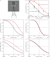
Figure 4.
Effect of intracortical microstimulation on judgements about a visual stimulus. Experimental data and simulations of psychometric functions illustrate different microstimulation effects and strategies that may occur. (a) Illustration of the visual cylinder task, in which monkeys discriminated the direction of rotation of a transparent structure-from-motion cylinder presented in the receptive field (white dashed circle) of microstimulation sites in extrastriate visual area V5/MT. The direction of rotation was defined by separating front and back surfaces with binocular disparity. The animal indicated its perceptual choice with an eye movement to one of two targets, located at opposite sides of the fixation point. Animals were rewarded for a correct choice with respect to the visual stimulus. (b) Gaussian psychometric functions (PMFs) fitted to experimental data from Krug et al. [88] with microstimulation at cortical site ica197, which was tuned for a negative cylinder disparity. Electrical microstimulation at this site induced a strong perceptual shift in the PMF in the preferred direction (PREF) of the neurons at the stimulation site in V5/MT. The animal's ‘null bias’ also caused the PMFs to shift towards the null direction (NULL), which is apparent in the non-stimulated trials (red lines: microstimulated trials; black lines: non-stimulated trials). Panels (cf) illustrate alternative possible outcomes based on data simulations. (c) Simulation of the PMFs that we would expect to see if the null bias were not present. The shift in the PMF due to electrical microstimulation would be the same, but the PMF for non-stimulated trials would pass through 50% at zero disparity. (d) Simulation for the hypothetical case where an animal could detect microstimulation trials and apply the null bias on microstimulation trials only. As in the discussed experiments, animals would be rewarded for correctly reporting the visual stimulus only. So we would expect that the perceptual shift due to microstimulation might be all but cancelled out by such a strategy. (e) Simulated experiment, in which microstimulation detection training (for microstimulation alone) at a direction- and disparity-selective V5/MT site is followed by microstimulation at the same site during the cylinder task. Expected PMF for the cylinder task is shown if microstimulation detection training simply increased visual discrimination thresholds, as reported in Ni & Maunsell [36], without affecting the integration of electrical and visual stimulation. The PMF flattens as performance accuracy decreases, but the bias effect due to microstimulation remains. (f) As for (e), but now the microstimulation perceptual shift is cancelled out because having been trained to detect microstimulation, animals might be able to distinguish microstimulation trials and apply the null bias on those trials only.

References

    1. Brodmann K. 1909. Vergleichende Lokalisationslehre der Grosshirnrinde in ihren Prinzipien dargestellt auf Grund des Zellenbaues. Leipzig, Germany: Johann Ambrosius Barth.
    1. Felleman DJ, Van Essen DC. 1991. Distributed hierarchical processing in the primate cerebral cortex. Cereb. Cortex 1, 1–47. (10.1093/cercor/1.1.1) - DOI - PubMed
    1. Logothetis NK, Pauls J, Augath M, Trinath T, Oeltermann A. 2001. Neurophysiological investigation of the basis of the fMRI signal. Nature 412, 150–157. (10.1038/35084005) - DOI - PubMed
    1. Logothetis NK, Wandell BA. 2004. Interpreting the BOLD signal. Annu. Rev. Physiol. 66, 735–769. (10.1146/annurev.physiol.66.082602.092845) - DOI - PubMed
    1. Yacoub E, et al. 2001. Imaging brain function in humans at 7 Tesla. Magn. Reson. Med. 45, 588–594. (10.1002/mrm.1080) - DOI - PubMed

Publication types

LinkOut - more resources