. 2015 Jun:34:65-86.
doi: 10.3767/003158515X685698. Epub 2014 Dec 1.

Application of the consolidated species concept to Cercospora spp. from Iran

Affiliations

Application of the consolidated species concept to Cercospora spp. from Iran

M Bakhshi et al. Persoonia. 2015 Jun.

Abstract

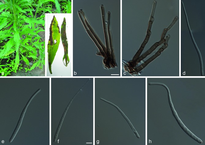
The genus Cercospora includes many important plant pathogenic fungi associated with leaf spot diseases on a wide range of hosts. The mainland of Iran covers various climatic regions with a great biodiversity of vascular plants, and a correspondingly high diversity of cercosporoid fungi. However, most of the cercosporoid species found to date have been identified on the basis of morphological characteristics and there are no cultures that support these identifications. In this study the Consolidated Species Concept was applied to differentiate Cercospora species collected from Iran. A total of 161 Cercospora isolates recovered from 74 host species in northern Iran were studied by molecular phylogenetic analysis. Our results revealed a rich diversity of Cercospora species in northern Iran. Twenty species were identified based on sequence data of five genomic loci (ITS, TEF1-α, actin, calmodulin and histone H3), host, cultural and morphological data. Six novel species, viz. C. convolvulicola, C. conyzae-canadensis, C. cylindracea, C. iranica, C. pseudochenopodii and C. sorghicola, are introduced. The most common taxon was Cercospora cf. flagellaris, which remains an unresolved species complex with a wide host range. New hosts were recorded for previously known Cercospora species, including C. apii, C. armoraciae, C. beticola, C. cf. richardiicola, C. rumicis, Cercospora sp. G and C. zebrina.

Keywords: Cercospora apii complex; Mycosphaerella; biodiversity; cercosporoid; host specificity; leaf spot; multilocus sequence typing (MLST); taxonomy.


Figures

Fig. 1
Fig. 1
Consensus phylogram (50 % majority rule) of 43 082 trees resulting from a Bayesian analysis of the combined 5-gene sequence alignment using MrBayes v. 3.2.1. Bayesian posterior probabilities are indicated with colour-coded branches and numbers (see legend) and the scale bar indicates 0.2 expected changes per site. Lineages from Iran are indicated in coloured blocks and species names in black text. Hosts and provinces of origin are indicated in green and brown text, respectively. The tree was rooted to Ramularia endophylla (isolate CBS 113265).
Fig. 1
Fig. 1
Consensus phylogram (50 % majority rule) of 43 082 trees resulting from a Bayesian analysis of the combined 5-gene sequence alignment using MrBayes v. 3.2.1. Bayesian posterior probabilities are indicated with colour-coded branches and numbers (see legend) and the scale bar indicates 0.2 expected changes per site. Lineages from Iran are indicated in coloured blocks and species names in black text. Hosts and provinces of origin are indicated in green and brown text, respectively. The tree was rooted to Ramularia endophylla (isolate CBS 113265).
Fig. 2
Fig. 2
Cercospora chenopodii (CCTU 1033). a. Leaf spots; b. c. fasciculate conidiophores; d–g. conidia. — Scale bars = 10 μm.
Fig. 3
Fig. 3
Cercospora convolvulicola (CBS 136126). a. Leaf spots; b. c. fasciculate conidiophores; d–j. conidia. — Scale bars = 10 μm.
Fig. 4
Fig. 4
Cercospora conyzae-canadensis (CBS 135978). a. Leaf spots; b. c. fasciculate conidiophores; d–h. conidia. — Scale bars = 10 μm.
Fig. 5
Fig. 5
Cercospora cylindracea (CBS 138580). a. Leaf spots on Cichorium intybus; b. leaf spots on Lactuca serriola; c–e. fasciculate conidiophores; f–k. conidia. — Scale bars = 10 μm.
Fig. 6
Fig. 6
Cercospora iranica (CBS 136124). a. Leaf spots; b. fasciculate conidiophores; c–h. conidia. — Scale bars = 10 μm.
Fig. 7
Fig. 7
Cercospora pseudochenopodii (CBS 136022). a. Leaf spots; b. c. fasciculate conidiophores; d–h. conidia. — Scale bars = 10 μm.
Fig. 8
Fig. 8
Cercospora solani (CBS 136038). a. Leaf spots; b. c. fasciculate conidiophores; d–h. conidia. — Scale bars = 10 μm.
Fig. 9
Fig. 9
Cercospora sorghicola (CBS 136448). a. Leaf spots; b. c. fasciculate conidiophores; d–h. conidia. — Scale bars = 10 μm.
Fig. 10
Fig. 10
Consensus phylogram (50 % majority rule) of 346 trees resulting from a Bayesian analysis of the ITS sequence alignment using MrBayes v. 3.2.1. The tree was rooted to Ramularia endophylla (strain CBS 113265).
Fig. 11
Fig. 11
Cercospora sp. T (CBS 136125). a. b. Leaf spots; c. intraepidermal caespituli; d. substomatal caespituli; e. fasciculate conidiophores; f–h. conidia. — Scale bars = 10 μm.

References

    1. Agrios GN. 2005. Plant pathology, fifth edition Academic Press, New York.
    1. Aptroot A. 2006. Mycosphaerella and its anamorphs: 2. Conspectus of Mycosphaerella. CBS Biodiversity Series 5: 1–231.
    1. Arzanlou M, Abeln EC, Kema GH, et al. 2007. Molecular diagnostics for the Sigatoka disease complex of banana. Phytopathology 97: 1112–1118. - PubMed
    1. Arzanlou M, Groenewald J, Fullerton R, et al. 2008. Multiple gene genealogies and phenotypic characters differentiate several novel species of Mycosphaerella and related anamorphs on banana. Persoonia 20: 19–37. - PMC - PubMed
    1. Bakhshi M, Arzanlou M, Babai-Ahari A. 2011. Uneven distribution of mating type alleles in Iranian populations of Cercospora beticola, the causal agent of Cercospora leaf spot disease of sugar beet. Phytopathologia Mediterranea 50: 101–109.

LinkOut - more resources