. 2015 Jul 28:4:384.
doi: 10.1186/s40064-015-1170-9. eCollection 2015.

Magnetic resonance imaging of uterine fibroids: a preliminary investigation into the usefulness of 3D-rendered images for surgical planning

Affiliations

Magnetic resonance imaging of uterine fibroids: a preliminary investigation into the usefulness of 3D-rendered images for surgical planning

Sayed Ahmad Zikri B Sayed Aluwee et al. Springerplus. .

Abstract

Purpose: This study aimed to assess the efficacy of 3D surface-rendered (SR) magnetic resonance (MR) images for surgical planning of uterine fibroids.

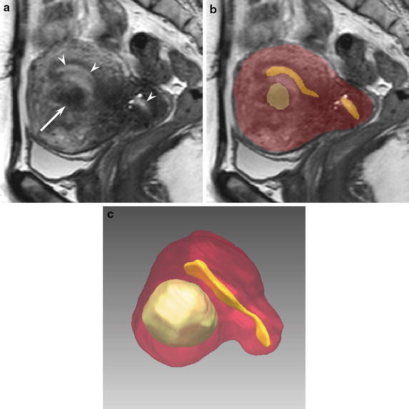
Methods: Ten patients with uterine fibroids underwent 3D volume isotropic turbo spin-echo acquisition (VISTA) sequences in sagittal planes. SR images showing the uterine body, endometrium, and fibroids were extracted from the raw MR data. The preoperative assessment for fertility-preserving fibroid enucleation was independently performed by two gynecologists using 2D sagittal and 3D SR images separately.

Results: The required interpretation times [second] for sagittal versus SR images were 19.7 ± 9.5 versus 10.4 ± 5.1 for observer 1 (p < 0.05) and 47.5 ± 12.3 versus 19.7 ± 9.5 for observer 2 (p < 0.01). The accuracy rates of the planned surgical procedures from sagittal versus SR images were 50 versus 70% for observer 1 and 70 versus 70% for observer 2. The accuracy rates of the numbers of fibroids to be removed from sagittal versus SR images were 70 versus 80% for observer 1 and 70 versus 80% for observer 2.

Conclusion: Compared with sagittal images, SR images could significantly reduce the time required for surgical planning of uterine fibroids without sacrificing the accuracy of the preoperative assessment.

Keywords: 3D; Fibroid; Magnetic resonance imaging; Uterus.


Figures

Fig. 1
Fig. 1
Process flow of the segmentation and 3D rendering. First, the target area (endometrium, fibroids, and uterine body) of the uterine MR images was segmented. Then, a correction process was applied to each slice from different image directions. Finally, a 3D surface rendering visualization was created.
Fig. 2
Fig. 2
A 54-year-old woman with uterine fibroids (Case 6). The gold standard for the planned surgical procedure was total laparoscopic myomectomy (TLM). a A sagittal 3D VISTA image (TR/TE, 1,800/205 ms) demonstrates hyperintensity in the endometrium (arrowheads) and hypointensity in a fibroid (arrow). b The segmentation results of the target regions indicate the uterine body as a red color, the endometrium as a yellow color, and fibroids as a green color. c A 3D SR image precisely indicates the positional relationship of the uterine body (red), endometrium (yellow), and fibroids (green).
Fig. 3
Fig. 3
A 39-year-old woman with uterine fibroids (Case 1). The gold standard for the planned surgical procedure was laparoscopically assisted myomectomy (LAM). a A Sagittal 3D VISTA image (TR/TE, 1,800/205 ms) indicates a subserosal fibroid (arrow) and intramural fibroids (arrowheads). b The segmentation results of the target regions indicate the uterine body as a red color, the endometrium as a yellow color, and fibroids as a green color. c A 3D SR image precisely indicates the positional relationship of the uterine body (red), endometrium (yellow), and fibroids (green).
Fig. 4
Fig. 4
Advanced visualization by combining 2D slice and 3D rendering views. a 2D cross-sectional views of uterine MR images provide the advantage of viewing 2D images in different planes. b A combination of 2D and 3D views to provide visualization of the uterine structures from different directions and with scaling. The positional relationship of the uterine structures with respect to others organ nearby and the types of fibroids can be understood easily with the combination.

References

    1. Berkeley AS, DeCherney AH, Polan ML. Abdominal myomectomy and subsequent fertility. Surg Gynecol Obstet. 1983;156(3):319–322. - PubMed
    1. Buttram VC, Jr, Reiter RC. Uterine leiomyomata: etiology, symptomatology, and management. Fertil Steril. 1981;36(4):433–445. - PubMed
    1. Dubuisson JB, Chapron C. Laparoscopic myomectomy today. A good technique when correctly indicated. Hum Reprod. 1996;11(5):934–935. doi: 10.1093/oxfordjournals.humrep.a019325. - DOI - PubMed
    1. Dubuisson JB, Chapron C, Chavet X, Gregorakis SS. Fertility after laparoscopic myomectomy of large intramural myomas: preliminary results. Hum Reprod. 1996;11(3):518–522. doi: 10.1093/HUMREP/11.3.518. - DOI - PubMed
    1. Fasih N, Prasad Shanbhogue AK, Macdonald DB, Fraser-Hill MA, Papadatos D, Kielar AZ, et al. Leiomyomas beyond the uterus: unusual locations, rare manifestations. Radiographics. 2008;28(7):1931–1948. doi: 10.1148/rg.287085095. - DOI - PubMed

LinkOut - more resources