. 2015:2015:248920.
doi: 10.1155/2015/248920. Epub 2015 Jul 9.

Tissue Remodelling following Resection of Porcine Liver

Affiliations

Tissue Remodelling following Resection of Porcine Liver

Ingvild Engdal Nygård et al. Biomed Res Int. 2015.

Abstract

Aim: To study genes regulating the extracellular matrix (ECM) and investigate the tissue remodelling following liver resection in porcine.

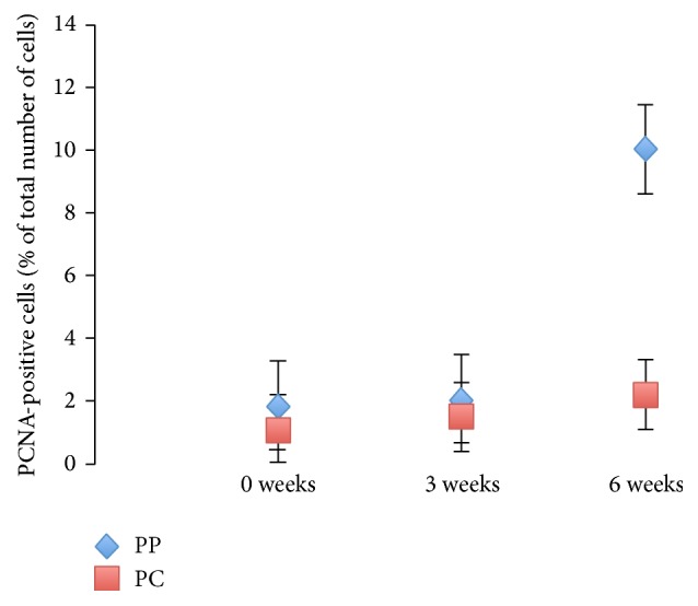
Methods: Four pigs with 60% partial hepatectomy- (PHx-) induced liver regeneration were studied over six weeks. Four pigs underwent sham surgery and another four pigs were used as controls of the normal liver growth. Liver biopsies were taken upon laparotomy, after three and six weeks. Gene expression profiles were obtained using porcine-specific oligonucleotide microarrays. Immunohistochemical staining was performed and a proliferative index was assessed.

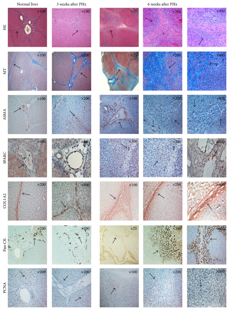
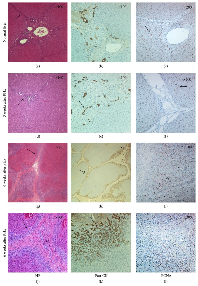
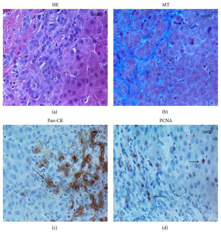
Results: More differentially expressed genes were associated with the regulation of ECM in the resection group compared to the sham and control groups. Secreted protein acidic and rich in cysteine (SPARC) and collagen 1, alpha 2 (COL1A2) were both upregulated in the early phase of liver regeneration, validated by immunopositive cells during the remodelling phase of liver regeneration. A broadened connective tissue was demonstrated by Masson's Trichrome staining, and an immunohistochemical staining against pan-Cytokeratin (pan-CK) demonstrated a distinct pattern of migrating cells, followed by proliferating cell nuclear antigen (PCNA) positive nuclei.

Conclusions: The present study demonstrates both a distinct pattern of PCNA positive nuclei and a deposition of ECM proteins in the remodelling phase of liver regeneration.


Figures

Figure 1
Figure 1
Overview of immunohistochemically stained normal and regenerated liver, three and six weeks after PHx. Staining for HE, Masson's Trichrome, α-SMA, SPARC, COL1A2, pan-CK and PCNA of a normal liver, a regenerated liver three weeks after PHx, and a regenerated liver six weeks after PHx, respectively. Original magnification at the upper right corner.
Figure 2
Figure 2
PCNA index. Percentage of PCNA-positive cells located in the periportal region (PP) and pericentral region (PC), at 0, 3, and 6 weeks after PHx.
Figure 3
Figure 3
HE, pan-CK, and PCNA staining throughout liver regeneration. (a, d, g, and j) HE staining of normal liver and regenerated liver. Note the broadening of the connective tissue at six weeks after PHx. (b, e, h and k) Pan-CK staining of normal and regenerated liver. Note the heavily stained cholangiocytes in the periportal region at six weeks after PHx. (c, f, i, and l) PCNA staining of normal and regenerated liver. Note the multiple cells immunopositive for PCNA presented along the connective tissue at six weeks after PHx. Original magnification at the upper right corner.
Figure 4
Figure 4
HE, MT, pan-CK, and PCNA at six weeks after PHx. (a and b) HE and Masson's Trichrome staining revealed hepatocyte-like cells located in between the fibres of the connective tissue. (c and d) Pan-CK and PCNA-positive cells located both in the connective tissue layer and in the adjacent hepatocyte parenchymal tissue. Original magnification at the upper right corner.

References

    1. Taub R. Liver regeneration: from myth to mechanism. Nature Reviews Molecular Cell Biology. 2004;5(10):836–847. doi: 10.1038/nrm1489. - DOI - PubMed
    1. Nygård I. E., Mortensen K. E., Hedegaard J., et al. The genetic regulation of the terminating phase of liver regeneration. Comparative Hepatology. 2012;11(1, article 3) doi: 10.1186/1476-5926-11-3. - DOI - PMC - PubMed
    1. Lin C.-W., Mars W. M., Paranjpe S., et al. Hepatocyte proliferation and hepatomegaly induced by phenobarbital and 1,4-bis [2-(3,5-dichloropyridyloxy)] benzene is suppressed in hepatocyte-targeted glypican 3 transgenic mice. Hepatology. 2011;54(2):620–630. doi: 10.1002/hep.24417. - DOI - PMC - PubMed
    1. Yuan X., Yan S., Zhao J., et al. Lipid metabolism and peroxisome proliferator-activated receptor signaling pathways participate in late-phase liver regeneration. Journal of Proteome Research. 2011;10(3):1179–1190. doi: 10.1021/pr100960h. - DOI - PubMed
    1. Chang C., Xu C. Transcriptome atlas of glutamine family amino acid metabolism-related genes in eight regenerating liver cell types. Cell Biology International. 2010;34(12):1189–1198. doi: 10.1042/CBI20090352. - DOI - PubMed

Publication types

Substances