

Synaptic connections of amacrine cells containing vesicular glutamate transporter 3 in baboon retinas
- PMID: 26241195
- PMCID: PMC4943031
- DOI: 10.1017/S0952523815000036
Synaptic connections of amacrine cells containing vesicular glutamate transporter 3 in baboon retinas
Abstract
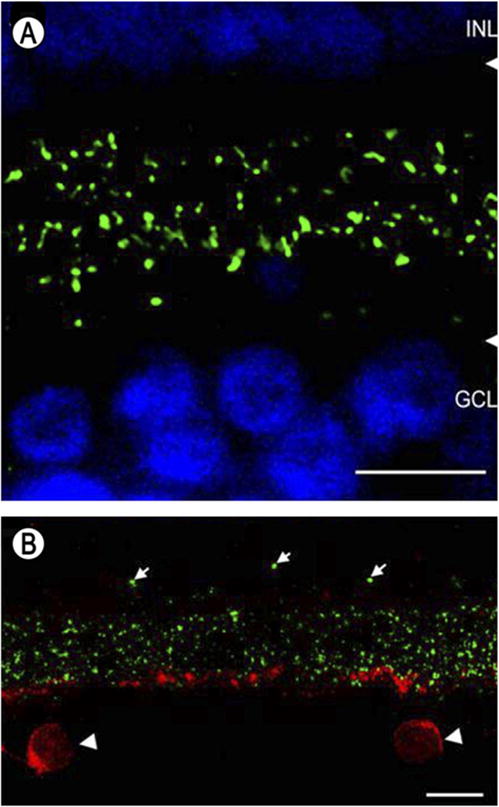
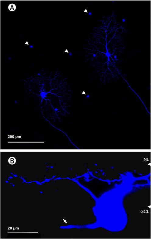
The goals of these experiments were to describe the morphology and synaptic connections of amacrine cells in the baboon retina that contain immunoreactive vesicular glutamate transporter 3 (vGluT3). These amacrine cells had the morphology characteristic of knotty bistratified type 1 cells, and their dendrites formed two plexuses on either side of the center of the inner plexiform layer. The primary dendrites received large synapses from amacrine cells, and the higher-order dendrites were both pre- and postsynaptic to other amacrine cells. Based on light microscopic immunolabeling results, these include AII cells and starburst cells, but not the polyaxonal amacrine cells tracer-coupled to ON parasol ganglion cells. The vGluT3 cells received input from ON bipolar cells at ribbon synapses and made synapses onto OFF bipolar cells, including the diffuse DB3a type. Many synapses from vGluT3 cells onto retinal ganglion cells were observed in both plexuses. At synapses where vGluT3 cells were presynaptic, two types of postsynaptic densities were observed; there were relatively thin ones characteristic of inhibitory synapses and relatively thick ones characteristic of excitatory synapses. In the light microscopic experiments with Neurobiotin-injected ganglion cells, vGluT3 cells made contacts with midget and parasol ganglion cells, including both ON and OFF types. Puncta containing immunoreactive gephyrin, an inhibitory synapse marker, were found at appositions between vGluT3 cells and each of the four types of labeled ganglion cells. The vGluT3 cells did not have detectable levels of immunoreactive γ-aminobutyric acid (GABA) or immunoreactive glycine transporter 1. Thus, the vGluT3 cells would be expected to have ON responses to light and make synapses onto neurons in both the ON and the OFF pathways. Taken with previous results, these findings suggest that vGluT3 cells release glycine at some of their output synapses and glutamate at others.
Keywords: Gephyrin; Glycine; Midget; Parasol; Primate.
Figures


Similar articles
-
Characterization of an amacrine cell type of the mammalian retina immunoreactive for vesicular glutamate transporter 3.J Comp Neurol. 2004 Jan 6;468(2):251-63. doi: 10.1002/cne.10962. J Comp Neurol. 2004. PMID: 14648683
-
Wavy multistratified amacrine cells in the monkey retina contain immunoreactive secretoneurin.Peptides. 2017 Aug;94:33-42. doi: 10.1016/j.peptides.2017.06.005. Epub 2017 Jun 19. Peptides. 2017. PMID: 28641988 Free PMC article.
-
Circuitry and role of substance P-immunoreactive neurons in the primate retina.J Comp Neurol. 1998 Apr 20;393(4):439-56. J Comp Neurol. 1998. PMID: 9550150
-
A tale of two neurotransmitters.Vis Neurosci. 2016 Jan;33:E017. doi: 10.1017/S0952523816000146. Vis Neurosci. 2016. PMID: 28359349 Free PMC article. Review.
-
On and off pathways through amacrine cells in mammalian retina: the synaptic connections of "starburst" amacrine cells.Vision Res. 1983;23(11):1265-79. doi: 10.1016/0042-6989(83)90102-5. Vision Res. 1983. PMID: 6362185 Review.
Cited by
-
Glutamate Cotransmission in Cholinergic, GABAergic and Monoamine Systems: Contrasts and Commonalities.Front Neural Circuits. 2018 Dec 18;12:113. doi: 10.3389/fncir.2018.00113. eCollection 2018. Front Neural Circuits. 2018. PMID: 30618649 Free PMC article. Review.
-
Functional Implications of Neurotransmitter Segregation.Front Neural Circuits. 2021 Oct 13;15:738516. doi: 10.3389/fncir.2021.738516. eCollection 2021. Front Neural Circuits. 2021. PMID: 34720888 Free PMC article. Review.
-
Melanopsin-mediated amplification of cone signals in the human visual cortex.Proc Biol Sci. 2024 May;291(2023):20232708. doi: 10.1098/rspb.2023.2708. Epub 2024 May 29. Proc Biol Sci. 2024. PMID: 38808443 Free PMC article.
-
Local synaptic integration enables ON-OFF asymmetric and layer-specific visual information processing in vGluT3 amacrine cell dendrites.Proc Natl Acad Sci U S A. 2017 Oct 24;114(43):11518-11523. doi: 10.1073/pnas.1711622114. Epub 2017 Sep 27. Proc Natl Acad Sci U S A. 2017. PMID: 28973895 Free PMC article.
-
Interactions between Glycine and Glutamate through Activation of Their Transporters in Hippocampal Nerve Terminals.Biomedicines. 2023 Nov 27;11(12):3152. doi: 10.3390/biomedicines11123152. Biomedicines. 2023. PMID: 38137373 Free PMC article.
References
-
- Calkins DJ, Schein SJ, Tsukamoto Y, Sterling P. M and L cones in macaque fovea connect to midget ganglion cells by different numbers of excitatory synapses. Nature. 1994;371:70–72. - PubMed
-
- Deniz S, Wersinger E, Schwab Y, Mura C, Erdelyi F, Szabo G, Rendon A, Sahel JA, Picaud S, Roux MJ. Mammalian retinal horizontal cells are unconventional GABAergic neurons. Journal of Neurochemistry. 2011;116:350–362. - PubMed
-
- Didier A, Carleton A, Bjaalie JG, Vincent JD, Ottersen OP, Storm-Mathisen J, Lledo PM. A dendrodendritic reciprocal synapse provides a recurrent excitatory connection in the olfactory bulb. Proceedings of the National Academy of Sciences of the United States of America. 2001;98:6441–6446. - PMC - PubMed
Publication types
MeSH terms
Substances
Grants and funding
LinkOut - more resources
Full Text Sources
Miscellaneous
