. 2015 Aug 4;5(8):e612.
doi: 10.1038/tp.2015.102.

Abnormal subcellular localization of GABAA receptor subunits in schizophrenia brain

Affiliations

Abnormal subcellular localization of GABAA receptor subunits in schizophrenia brain

T M Mueller et al. Transl Psychiatry. .

Abstract

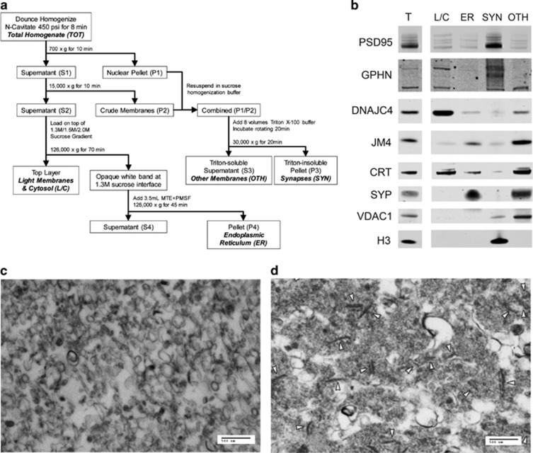
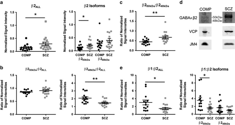
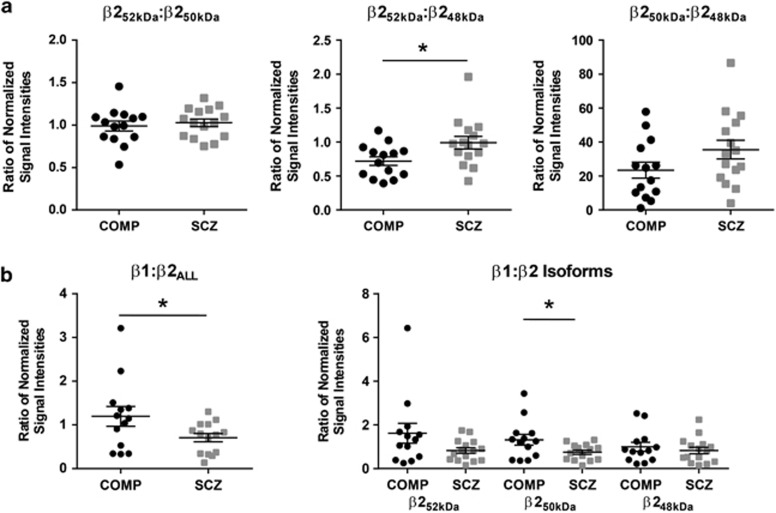
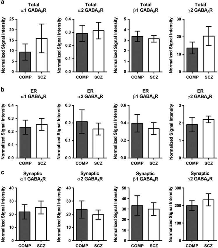
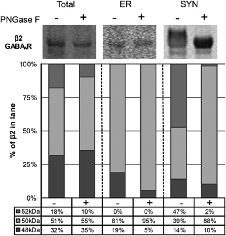
Inhibitory neurotransmission is primarily mediated by γ-aminobutyric acid (GABA) activating synaptic GABA type A receptors (GABA(A)R). In schizophrenia, presynaptic GABAergic signaling deficits are among the most replicated findings; however, postsynaptic GABAergic deficits are less well characterized. Our lab has previously demonstrated that although there is no difference in total protein expression of the α1-6, β1-3 or γ2 GABA(A)R subunits in the superior temporal gyrus (STG) in schizophrenia, the α1, β1 and β2 GABA(A)R subunits are abnormally N-glycosylated. N-glycosylation is a posttranslational modification that has important functional roles in protein folding, multimer assembly and forward trafficking. To investigate the impact that altered N-glycosylation has on the assembly and trafficking of GABA(A)Rs in schizophrenia, this study used western blot analysis to measure the expression of α1, α2, β1, β2 and γ2 GABA(A)R subunits in subcellular fractions enriched for endoplasmic reticulum (ER) and synapses (SYN) from STG of schizophrenia (N = 16) and comparison (N = 14) subjects and found evidence of abnormal localization of the β1 and β2 GABA(A)R subunits and subunit isoforms in schizophrenia. The β2 subunit is expressed as three isoforms at 52 kDa (β2(52 kDa)), 50 kDa (β2(50 kDa)) and 48 kDa (β2(48 kDa)). In the ER, we found increased total β2 GABA(A)R subunit (β2(ALL)) expression driven by increased β2(50 kDa), a decreased ratio of β(248 kDa):β2(ALL) and an increased ratio of β2(50 kDa):β2(48 kDa). Decreased ratios of β1:β2(ALL) and β1:β2(50 kDa) in both the ER and SYN fractions and an increased ratio of β2(52 kDa):β(248 kDa) at the synapse were also identified in schizophrenia. Taken together, these findings provide evidence that alterations of N-glycosylation may contribute to GABAergic signaling deficits in schizophrenia by disrupting the assembly and trafficking of GABA(A)Rs.


Figures

Figure 1
Figure 1
Fractions enriched for light membranes and cytosol (L/C), endoplasmic reticulum (ER), excitatory and inhibitory synapses (SYN) and other intermediate membranes (OTH) generated from postmortem human cortex. (a) Schematic depiction of the centrifugation, differential sucrose gradient and Triton solubilization steps to produce L/C, ER, SYN and OTH-enriched fractions from nitrogen-cavitated samples. Briefly, after nitrogen-cavitated cortical homogenate samples undergo sequential centrifugations, S2 is loaded on top of a differential sucrose gradient and ultracentrifuged to separate the ER from other light membranes and cytosolic components based on membrane density. The ER membranes appear as a semi-opaque white band, and the L/C remains suspended in the translucent top layer. P1 and P2 from the first centrifugation steps are resuspended, combined and solubilized by a brief incubation with Triton X-100 buffer. Following centrifugation, the Triton-insoluble synaptic membranes are concentrated in the resulting P3, while S3 contains the remaining heavy and intermediate membrane components. (b) Representative images from western blots of total homogenate, L/C, ER, SYN and OTH fractions probed for various subcellular markers to validate the efficacy of the fractionation method in postmortem human cortex. Target marker proteins include postsynaptic density protein 95 (PSD95), for excitatory synapses; gephyrin (GPHN), for inhibitory synapses and extrasynaptic membrane; DnaJ/hsp40 homolog subfamily C member 4 (DNAJC4), for cytosol; PRA1 family protein 2 (JM4), for ER and Golgi membranes; calreticulin (CRT), for ER lumen; synaptophysin (SYP), for extrasynaptic membranes; voltage dependent anion-selective channel protein 1 (VDAC), for mitochondria; and histone 3 (H3), for nuclei. (c and d) Representative electron microscopy (EM) image of the ER and SYN fractions (scale bars, 500 nm). (c) ER membrane is enriched and no other identifiable structures or organelles are evident in the ER fraction. (d) Synapses, indicated by white arrowheads, are enriched and no other intact structures or organelles are visualized in the SYN fraction. MTE, D-mannitol, Tris-base, and EDTA; PMSF, phenylmethylsulfonyl fluoride.
Figure 2
Figure 2
The β2 GABAAR subunit is abnormally expressed in an isoform-specific manner and the ratios of β1 and β2 subunit isoforms are altered in the ER in schizophrenia. Western blot analysis of total β2 GABAAR subunit (β2ALL) and individual β2 GABAAR subunit 50 kDa and 48 kDa isoforms (β250 kDa and β248 kDa, respectively), the ratios of β2 GABAAR subunit isoforms to each other, and the ratio of β1:β2 GABAAR subunit and subunit isoform expression in the ER fraction in schizophrenia and comparison subjects. (a) ER fraction-normalized expression of β2ALL, and specifically the β250 kDa GABAAR subunit isoform, is increased in schizophrenia. (b) The ratio of β248 kDa:β2ALL GABAAR subunit fraction-normalized expression is decreased in the ER in schizophrenia. (c) The ratio of β250 kDa:β248 kDa GABAAR subunit fraction-normalized signal intensities is increased in schizophrenia. (d) Representative images of western blots of the β2 GABAAR subunit, VCP and JM4 from the ER fraction from comparison and schizophrenia subjects with the β250 kDa and β248 kDa protein bands indicated. (e) The ratio of β1:β2ALL, and specifically the ratio of β1:β250 kDa GABAAR subunit expression is significantly less in the ER fraction in schizophrenia. Data are expressed as either the signal intensity of protein targets in the ER fraction normalized to VCP as a loading control and JM4 as an ER marker relative to the VCP-normalized signal intensity of the same target in the total homogenate, or expressed as a ratio of normalized signal intensities, for each data point with means±s.e.m. for each group indicated in a, b, c and e. *P<0.05, **P<0.01. COMP, comparison subject; ER, endoplasmic reticulum; GABAAR, γ-aminobutyric acid type A receptor; SCZ, schizophrenia; VCP, valosin-containing protein.
Figure 3
Figure 3
The ratio of β1:β2ALL, β1:β250 kDa and β252 kDa:β248 kDa GABAAR subunit expression is increased in the SYN fraction in schizophrenia. Western blot analysis of the ratios of β1 and β2 GABAAR subunit isoform expression in the SYN fraction in schizophrenia and comparison subjects. (a) The ratio of β252 kDa:β248 kDa GABAAR subunit expression is increased in schizophrenia, with no difference between groups for the ratio of β252 kDa:β250 kDa or β250 kDa:β248 kDa GABAAR subunit expression in the SYN fraction. (b) The ratio of β1:β2ALL and β1:β250 kDa GABAAR subunit expression is decreased in the SYN fraction in schizophrenia. Data are expressed as a ratio of the signal intensity of protein targets in the SYN fraction normalized to VCP as a loading control and gephyrin as an inhibitory synaptic marker relative to the VCP-normalized signal intensity of the same targets in total homogenate for each subject; data are means±s.e.m. *P<0.05. COMP, comparison subject; GABAAR, γ-aminobutyric acid type A receptor; SCZ, schizophrenia; SYN, synapse; VCP, valosin-containing protein.
Figure 4
Figure 4
α1, α2, β1 and γ2 GABAAR subunit expression are not different between diagnostic groups in the total homogenate, ER or SYN fractions. Western blot analysis of α1, α2ALL, β1 and γ2 GABAAR subunit expression in schizophrenia and comparison subjects. There are no differences between diagnostic groups in the protein expression of α1, α2ALL, β1 or γ2 GABAAR subunits in (a) the total homogenates, (b) ER fractions or (c) the SYN fractions. Data are expressed as the mean signal intensity (±s.e.m.) of protein targets in the ER fraction normalized to VCP as a loading control, and JM4 as an ER marker or gephyrin as an inhibitory synapse marker, relative to the VCP-normalized signal intensity of the same target in the total homogenate. COMP, comparison subject; ER, endoplasmic reticulum; GABAAR, γ-aminobutyric acid type A receptor; SCZ, schizophrenia; SYN, synapse; VCP, valosin-containing protein.
Figure 5
Figure 5
The β252 kDa GABAAR subunit isoform is N-glycosylated in postmortem human cortex. Representative images of western blots probed for the β2 GABAAR subunit in total homogenate, ER and SYN fractions with and without N-glycans cleaved by treatment with the deglycosylating enzyme PNGase F and corresponding graphs of β2 GABAAR subunit isoform protein expression as a percentage of total β2 GABAAR subunit in each lane. In brief, subcellular fractions generated from cortical homogenates were denatured and deglycosylated with PNGase F. Image Studio software was used to measure the signal intensity of protein bands at 52, 50 and 48 kDa in each lane. The signal intensity of each isoform was then divided by the sum of signal intensities for all the three isoforms to determine the percentage of total β2 GABAAR subunit expressed in the lane. After deglycosylation with PNGase F, the percentage of β252 kDa GABAAR is greatly reduced with a corresponding increase of β250 kDa GABAAR expressed in the SYN fraction. The calculated percentage of β248 kDa GABAAR in the ER fraction is also reduced after PNGase F treatment; however, this is likely an artifact due to the low signal intensity values for protein bands measured in those lanes. ER, endoplasmic reticulum; GABAAR, γ-aminobutyric acid type A receptor; PNGase F, peptide N-glycosidase F; SCZ, schizophrenia; SYN, synapse.

Similar articles

Cited by

References

    1. Gonzalez-Burgos G, Fish KN, Lewis DA. GABA neuron alterations, cortical circuit dysfunction and cognitive deficits in schizophrenia. Neural Plast. 2011;2011:723184. - PMC - PubMed
    1. Lewis DA, Hashimoto T, Volk DW. Cortical inhibitory neurons and schizophrenia. Nat Rev Neurosci. 2005;6:312–324. - PubMed
    1. Lewis DA, Curley AA, Glausier JR, Volk DW. Cortical parvalbumin interneurons and cognitive dysfunction in schizophrenia. Trends Neurosci. 2012;35:57–67. - PMC - PubMed
    1. Uhlhaas PJ, Singer W. Neural synchrony in brain disorders: relevance for cognitive dysfunctions and pathophysiology. Neuron. 2006;52:155–168. - PubMed
    1. Nakazawa K, Zsiros V, Jiang Z, Nakao K, Kolata S, Zhang S, et al. GABAergic interneuron origin of schizophrenia pathophysiology. Neuropharmacology. 2012;62:1574–1583. - PMC - PubMed

Publication types

MeSH terms