Increased nuclear β-catenin expression in oral potentially malignant lesions: A marker of epithelial dysplasia
- PMID: 26241451
- PMCID: PMC4598921
- DOI: 10.4317/medoral.20341
Increased nuclear β-catenin expression in oral potentially malignant lesions: A marker of epithelial dysplasia
Abstract
Background: Deregulation of β-catenin is associated with malignant transformation; however, its relationship with potentially malignant and malignant oral processes is not fully understood. The aim of this study was to determine and compare the nuclear β-catenin expression in oral dysplasia and oral squamous cell carcinoma (OSCC).
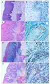
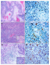
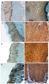
Material and methods: Cross sectional study. Immunodetection of β-catenin was performed on 72 samples, with the following distribution: 21 mild dysplasia, 12 moderate dysplasia, severe dysplasia 3, 36 OSCC including 19 well differentiated, 15 moderately differentiated and 2 poorly differentiated. Through microscopic observation the number of positive cells per 1000 epithelial cells was counted. For the statistical analysis, the Kruskal Wallis test was used.
Results: Nuclear expression of β-catenin was observed in all samples with severe and moderate dysplasia, with a median of 267.5, in comparison to mild dysplasia whose median was 103.75. Only 10 samples (27.7%) with OSCC showed nuclear expression, with statistically significant differences between groups (p < 0.05).
Conclusions: Our results are consistent with most of the reports which show increased presence of β-catenin in severe and moderate dysplasia compared to mild dysplasia; however the expression of nuclear β-catenin decreased after starting the invasive neoplastic process. This suggests a role for this protein in the progression of dysplasia and early malignant transformation to OSCC. Immunodetection of β-catenin could be a possible immune marker in the detection of oral dysplasia.
Conflict of interest statement
Figures


Similar articles
-
beta- and gamma-catenin expression in oral dysplasia.Oral Oncol. 2009 Jun;45(6):501-4. doi: 10.1016/j.oraloncology.2008.06.004. Epub 2008 Aug 19. Oral Oncol. 2009. PMID: 18715817
-
Loss of ELF3 immunoexpression is useful for detecting oral squamous cell carcinoma but not for distinguishing between grades of epithelial dysplasia.Ann Diagn Pathol. 2013 Aug;17(4):331-40. doi: 10.1016/j.anndiagpath.2013.03.003. Epub 2013 May 3. Ann Diagn Pathol. 2013. PMID: 23643910
-
p73 expression for human buccal epithelial dysplasia and squamous cell carcinoma: does it correlate with nodal status of carcinoma and is there a relationship with malignant change of epithelial dysplasia?Head Neck. 2004 Nov;26(11):945-52. doi: 10.1002/hed.20098. Head Neck. 2004. PMID: 15390192
-
Wnt/β-Catenin Signaling in Oral Carcinogenesis.Int J Mol Sci. 2020 Jun 30;21(13):4682. doi: 10.3390/ijms21134682. Int J Mol Sci. 2020. PMID: 32630122 Free PMC article. Review.
-
Histological features of differentiated dysplasia in the oral mucosa: a review of oral invasive squamous cell carcinoma cases diagnosed with benign or low-grade dysplasia on previous biopsies.Hum Pathol. 2022 Aug;126:45-54. doi: 10.1016/j.humpath.2022.05.010. Epub 2022 May 18. Hum Pathol. 2022. PMID: 35597368 Review.
Cited by
-
The Role of Carcinogenesis-Related Biomarkers in the Wnt Pathway and Their Effects on Epithelial-Mesenchymal Transition (EMT) in Oral Squamous Cell Carcinoma.Cancers (Basel). 2020 Feb 28;12(3):555. doi: 10.3390/cancers12030555. Cancers (Basel). 2020. PMID: 32121061 Free PMC article. Review.
-
Immunohistochemical Assessment of Maspin, β-Catenin, and MMP-14 in Oral Potentially Malignant Lesions and Oral Squamous Cell Carcinoma: A Retrospective Observational Study.Medicina (Kaunas). 2025 Jun 4;61(6):1037. doi: 10.3390/medicina61061037. Medicina (Kaunas). 2025. PMID: 40572725 Free PMC article.
-
Exceptional Evolution of a Squamous Odontogenic Tumor in the Jaw: Molecular Approach.Int J Mol Sci. 2024 Sep 2;25(17):9547. doi: 10.3390/ijms25179547. Int J Mol Sci. 2024. PMID: 39273494 Free PMC article.
-
Expression profile of components of the β-catenin destruction complex in oral dysplasia and oral cancer.Med Oral Patol Oral Cir Bucal. 2021 Nov 1;26(6):e729-e737. doi: 10.4317/medoral.24528. Med Oral Patol Oral Cir Bucal. 2021. PMID: 34564680 Free PMC article.
-
Biomarkers of epithelial-mesenchymal transition: E-cadherin and beta-catenin in malignant transformation of oral lesions.Can J Dent Hyg. 2024 Jun 1;58(2):111-119. eCollection 2024 Jun. Can J Dent Hyg. 2024. PMID: 38974823 Free PMC article. Review.
References
-
- Warnakulasuriya S, Johnson NW, van der Waal I. Nomenclature and classification of potentially malignant disorders of the oral mucosa. J Oral Pathol Med. 2007;36:575–80. - PubMed
-
- Warnakulasuriya S, Reibel J, Bouquot J, Dabelsteen E. Oral epithelial dysplasia classification systems: predictive value, utility, weaknesses and scope for improvement. J Oral Pathol Med. 2008;37:127–33. - PubMed
-
- Reichart PA, Philipsen HP. Oral erythroplakia--a review. Oral Oncol. 2005;41:551–61. - PubMed
-
- Neville BW, Day TA. Oral cancer and precancerous lesions. CA Cancer J Clin. 2002;52:195–215. - PubMed
-
- Hall GL, Shaw RJ, Field EA, Rogers SN, Sutton DN, Woolgar JA. p16 Promoter methylation is a potential predictor of malignant transformation in oral epithelial dysplasia. Cancer Epidemiol Biomarkers Prev. 2008;17:2174–9. - PubMed
MeSH terms
Substances
LinkOut - more resources
Full Text Sources
Other Literature Sources
Medical

