

Differential Effects of Dabigatran and Warfarin on Bone Volume and Structure in Rats with Normal Renal Function
- PMID: 26241483
- PMCID: PMC4524674
- DOI: 10.1371/journal.pone.0133847
Differential Effects of Dabigatran and Warfarin on Bone Volume and Structure in Rats with Normal Renal Function
Abstract
Background: Warfarin, a widely used anticoagulant, is a vitamin K antagonist impairing the activity of vitamin K-dependent Bone Gla Protein (BGP or Osteocalcin) and Matrix Gla Protein (MGP). Because dabigatran, a new anticoagulant, has no effect on vitamin K metabolism, the aim of this study was to compare the impact of warfarin and dabigatran administration on bone structure and vascular calcification.
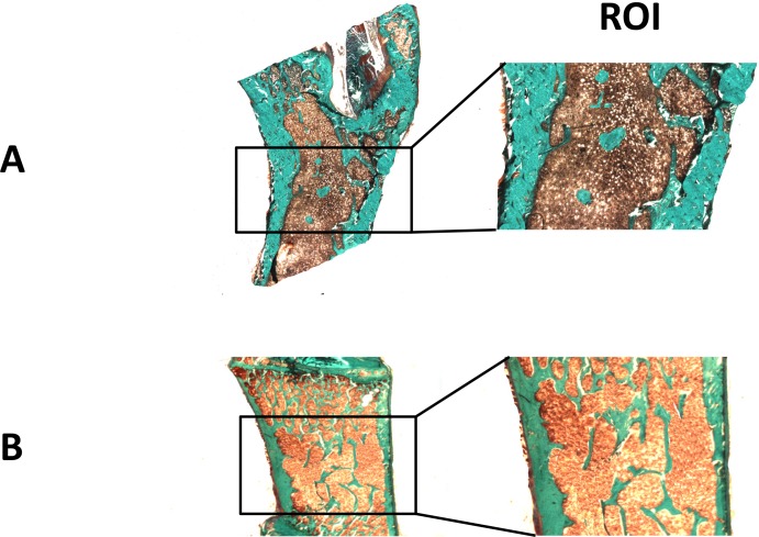
Methods: Rats with normal renal function received for 6 weeks warfarin, dabigatran or placebo. Bone was evaluated immuno-histochemically and hystomorphometrically after double labelling with declomycin and calcein. Aorta and iliac arteries were examined histologically.
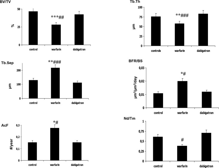
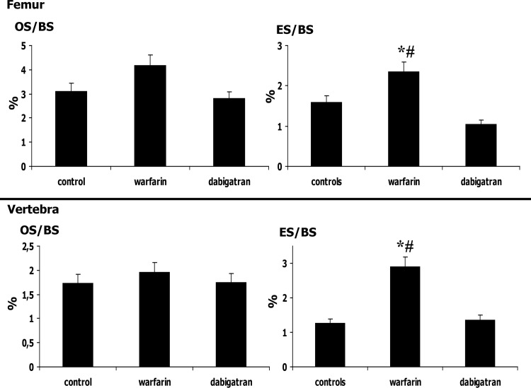
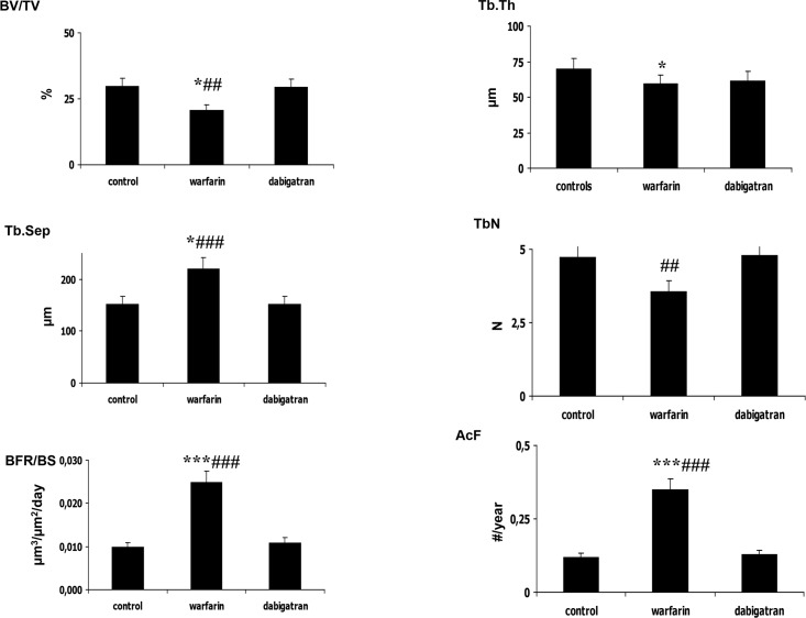
Results: Histomorphometric analysis of femur and vertebrae showed significantly decreased bone volume and increased trabecular separation in rats treated with warfarin. Vertebra analysis showed that the trabecular number was higher in dabigatran treated rats. Osteoblast activity and resorption parameters were similar among groups, except for maximum erosion depth, which was higher in warfarin treated rats, suggesting a higher osteoclastic activity. Therefore, warfarin treatment was also associated with higher bone formation rate/bone surface and activation frequency. Warfarin treatment may cause an increased bone turnover characterized by increased remodelling cycles, with stronger osteoclast activity compared to the other groups. There were no differences among experimental groups in calcium deposition either in aortic or iliac arteries.
Conclusions: These findings suggest for the first time that dabigatran has a better bone safety profile than warfarin, as warfarin treatment affects bone by reducing trabecular size and structure, increasing turnover and reducing mineralization. These differences could potentially result in a lower incidence of fractures in dabigatran treated patients.
Conflict of interest statement
Figures


Similar articles
-
Osteopenia and bone-remodeling abnormalities in warfarin-treated lambs.J Bone Miner Res. 1993 Dec;8(12):1417-26. doi: 10.1002/jbmr.5650081202. J Bone Miner Res. 1993. PMID: 8304041
-
Warfarin-induced artery calcification is accelerated by growth and vitamin D.Arterioscler Thromb Vasc Biol. 2000 Feb;20(2):317-27. doi: 10.1161/01.atv.20.2.317. Arterioscler Thromb Vasc Biol. 2000. PMID: 10669626
-
Long-term treatment with sodium warfarin results in decreased femoral bone strength and cancellous bone volume in rats.Thromb Res. 2002 Feb 15;105(4):353-8. doi: 10.1016/s0049-3848(02)00035-x. Thromb Res. 2002. PMID: 12031831
-
Bleeding, vertebral fractures and vascular calcifications in patients treated with warfarin: hope for lower risks with alternative therapies.Curr Vasc Pharmacol. 2011 Nov;9(6):763-9. doi: 10.2174/157016111797484134. Curr Vasc Pharmacol. 2011. PMID: 21623708 Review.
-
Effects of warfarin on biological processes other than haemostasis: A review.Food Chem Toxicol. 2018 Mar;113:19-32. doi: 10.1016/j.fct.2018.01.019. Epub 2018 Jan 17. Food Chem Toxicol. 2018. PMID: 29353071 Review.
Cited by
-
Comparative Risks of Fracture Among Direct Oral Anticoagulants and Warfarin: A Systematic Review and Network Meta-Analysis.Front Cardiovasc Med. 2022 May 23;9:896952. doi: 10.3389/fcvm.2022.896952. eCollection 2022. Front Cardiovasc Med. 2022. PMID: 35677694 Free PMC article.
-
Vitamin K and Bone Health: A Review on the Effects of Vitamin K Deficiency and Supplementation and the Effect of Non-Vitamin K Antagonist Oral Anticoagulants on Different Bone Parameters.J Osteoporos. 2019 Dec 31;2019:2069176. doi: 10.1155/2019/2069176. eCollection 2019. J Osteoporos. 2019. PMID: 31976057 Free PMC article. Review.
-
The Role of Vitamin K in CKD-MBD.Curr Osteoporos Rep. 2022 Feb;20(1):65-77. doi: 10.1007/s11914-022-00716-z. Epub 2022 Feb 8. Curr Osteoporos Rep. 2022. PMID: 35132525 Free PMC article. Review.
-
Drug-induced Reduction of Gamma Carboxylation in Osteocalcin: What is the Fallback?Cureus. 2019 Aug 28;11(8):e5504. doi: 10.7759/cureus.5504. Cureus. 2019. PMID: 31667038 Free PMC article. Review.
-
A Network Meta-Analysis Comparing Osteoporotic Fracture among Different Direct Oral Anticoagulants and Vitamin K Antagonists in Patients with Atrial Fibrillation.J Bone Metab. 2021 May;28(2):139-150. doi: 10.11005/jbm.2021.28.2.139. Epub 2021 May 31. J Bone Metab. 2021. PMID: 34130366 Free PMC article.
References
-
- Ansell J, Hirsh J, Hylek E, Jacobson A, Crowther M, Palareti G, et al. Pharmacology and management of the vitamin K antagonists: American College of Chest Physicians evidence-based clinical practice guidelines (8th Edition). Chest 2008; 133 (Suppl. 6): 160S–198S. - PubMed
-
- Luo G, Ducy P, McKee MD, Pinero GJ, Loyer E, Behringer RR, et al. Spontaneous calcification of arteries and cartilage in mice lacking matrix GLA protein. Nature. 1997; 386: 78–81. - PubMed
-
- Price PA, Faus SA, Williamson MK. Warfarin causes rapid calcification of the elastic lamellae in rat arteries and heart valves. Arterioscler Thromb Vasc Biol. 1998; 18: 1400–1407. - PubMed
-
- Weber P. Vitamin K and bone health. Nutrition. 2001; 17 (10): 880–887. - PubMed
Publication types
MeSH terms
Substances
LinkOut - more resources
Full Text Sources
Other Literature Sources
Medical
Miscellaneous
