. 2015 Sep:86:199-207.
doi: 10.1016/j.yjmcc.2015.07.021. Epub 2015 Aug 1.

Hematopoietic progenitors are required for proper development of coronary vasculature

Affiliations

Hematopoietic progenitors are required for proper development of coronary vasculature

Gentian Lluri et al. J Mol Cell Cardiol. 2015 Sep.

Abstract

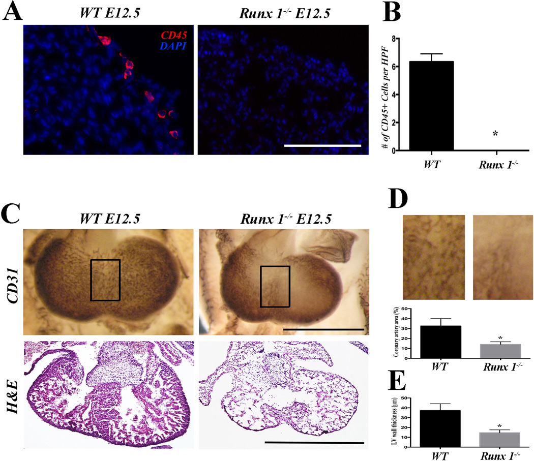
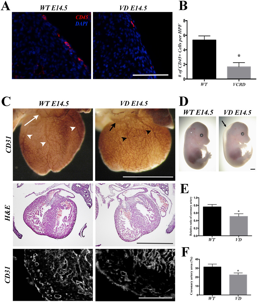
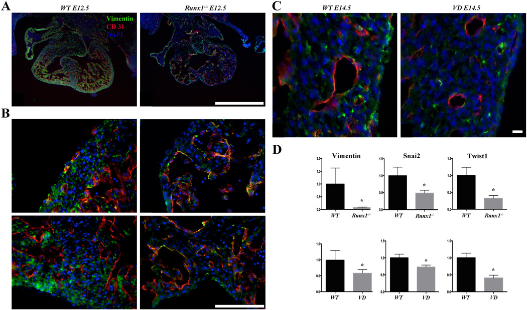
Rationale: During embryogenesis, hematopoietic cells appear in the myocardium prior to the initiation of coronary formation. However, their role is unknown.

Objective: Here we investigate whether pre-existing hematopoietic cells are required for the formation of coronary vasculature.

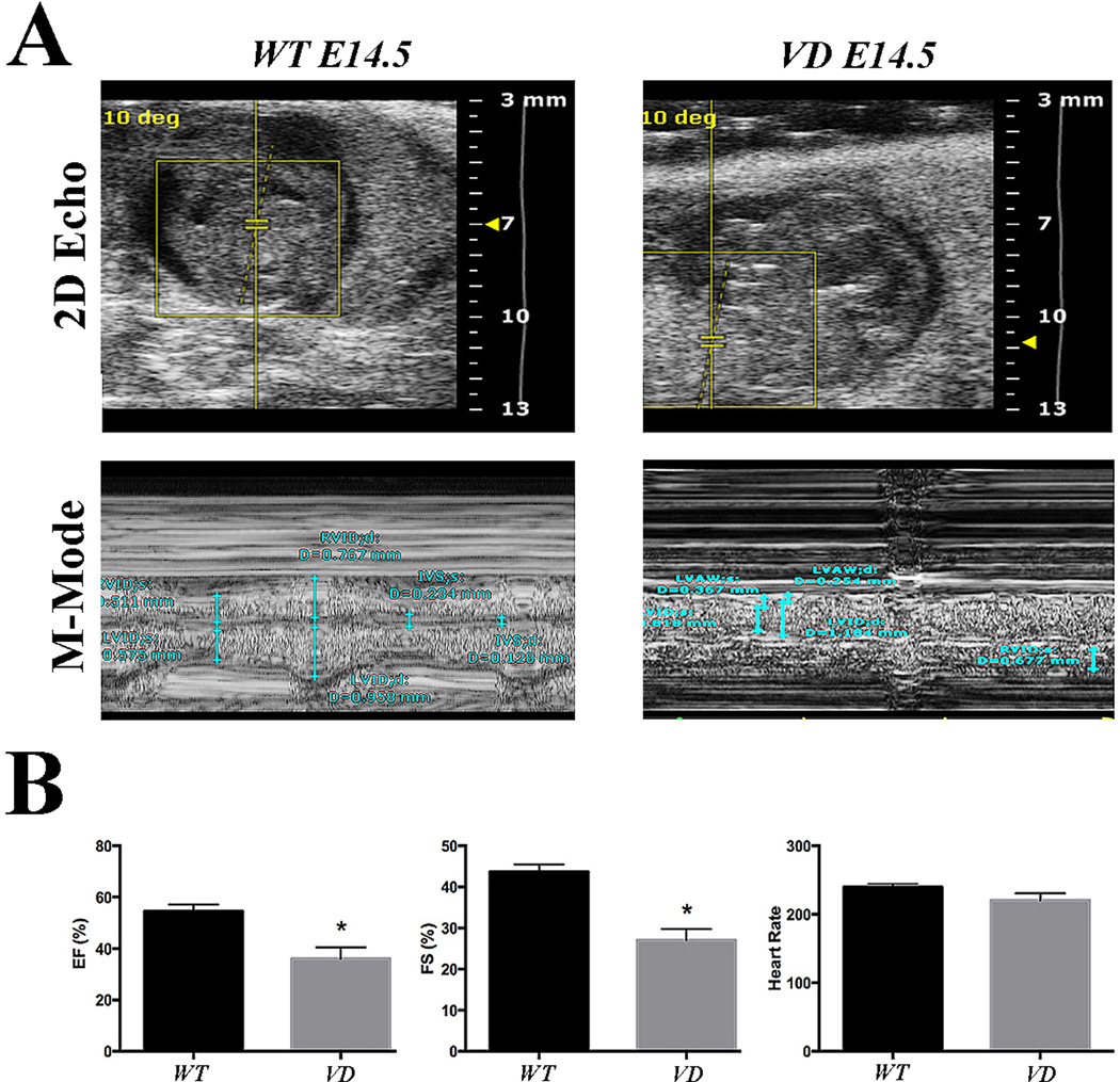
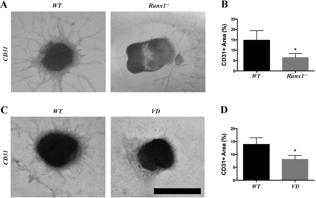
Methods and results: As a model of for hematopoietic cell deficient animals, we used Runx1 knockout embryos and Vav1-cre; R26-DTA embryos, latter of which genetically ablates 2/3 of CD45(+) hematopoietic cells. Both Runx1 knockout embryos and Vav1-cre; R26-DTA embryos revealed disorganized, hypoplastic microvasculature of coronary vessels on section and whole-mount stainings. Furthermore, coronary explant experiments showed that the mouse heart explants from Runx1 and Vav1-cre; R26-DTA embryos exhibited impaired coronary formation ex vivo. Interestingly, in both models it appears that epicardial to mesenchymal transition is adversely affected in the absence of hematopoietic progenitors.

Conclusion: Hematopoietic cells are not merely passively transported via coronary vessel, but substantially involved in the induction of the coronary growth. Our findings suggest a novel mechanism of coronary growth.

Keywords: Cardiac development; Coronary formation; EMT; Epicardium; Hematopoietic progenitors.


Conflict of interest statement

Disclosures and conflicts of interest: None

Figures

Figure 1
Figure 1. Runx1 mutant embryos show defects in cardiac development and coronary formation
(A) Representative CD45 staining of E12.5 Runx1-null embryos. No CD45+ cells are identified in the mutants. (B) Quantification of the number of CD45+ cells per high power field section (n=6). (C) Whole mount CD31 staining and H&E staining of E12.5 hearts. Coronary plexus formation is adversely affected in Runx1 null embryos (first row) when compared to wild-type embryos. H&E staining shows thin myocardium and ventricular septal defect (second row). (D) Quantification of the coronary plexus in the interventricular surface (boxed area in C; n=6). The density of the coronary plexus is reduced in Runx1 mutants. (E) Quantification of the thickness of ventricular free wall (n=6). Mutants show significantly thinner myocardium. Scale bars: A, 0.1 mm. C, 1 mm. * p < 0.05.
Figure 2
Figure 2. VD embryos shows abnormal hypoplastic coronary vessels and structural heart defects
(A) Representative CD45 staining of E14.5 VD embryos. Markedly reduced CD45+ cells are identified in the treated embryos. (B) Quantification of the number of the CD45+ cells per high power field section (n=5). (C) Whole mount CD31 staining, H&E staining, and section CD31 immunofluorescence staining of E14.5 hearts. Coronary plexus and major coronary vessels are adversely affected in the VD embryos (first row) when compared to wild-type embryos. H&E staining shows intact ventricular septum and myocardium (second row). Section CD31 immunostaining shows smaller coronary vessels (bottom row) in the VD embryos. (D) Representative E14.5 VD and wild-type embryos. VD embryos are of comparable size as wild-type embryos but exhibit subcutaneous edema (arrow). (E) Quantification of the relative length of the major coronary vessels in the wild-type hearts (white arrowheads in C) and VD hearts (black arrowheads in C) (n=5). (F) Quantification of the coronary plexus. The density of the coronary plexus is reduced in VD embryos (n=5). Scale bars: A, 0.1 mm. C (first and second row), 2 mm. C (bottom row), 0.1 mm. * p < 0.05.
Figure 3
Figure 3. VD embryos reveal reduced cardiac function
(A) Representative 2D images and M-mode analysis of E14.5 embryos. VD hearts show reduced systolic function (n=4). (B) Quantification of the ejection fraction (EF), fractional shortening (FS) and heart rate. The EF and FS are significantly reduced in the VD embryonic hearts (n=4). *p < 0.05
Figure 4
Figure 4. Runx1 and VD heart explants show reduced endothelial sprouting
(A) Representative ventricular explants from Runx1 mutant embryos. Sprouting of CD31+ cells is reduced in the mutants. (B) Quantification of the CD31+ sprouting area from Runx1 mutants (n=6). (C) Representative ventricular explants from VD embryos. CD31+ sprouting is reduced in the VD explants. (D) Quantification of the CD31+ sprouting area from VD explants (n=5). Scale bar: 1 mm. * p < 0.05.
Figure 5
Figure 5. Hematopoietic deficient embryos show EMT defect
(A) Representative vimentin (green) and CD31 (red) staining of E12.5 Runx1 mutant embryos. Markedly reduced vimentin staining is observed in the mutants (n=5). (B) High magnification of vimentin (green) and CD31 (red) staining of E12.5 Runx1 mutant embryos. The reduction of vimentin is observed throughout, including the lateral ventricular wall and interventricular septum as well (n=5). (C) Representative vimentin (green) and CD31 (red) staining of E14.5 VD embryos The reduction of vimentin is observed in VD hearts and it is associated with smaller size CD31+ vessels (n=4). (D) qRT-PCR of vimentin, snail2, and twist1. mRNA levels of vimentin, snail2, and twist1 are markedly downregulated in the epicardial cells of the E12.5 Runx1 mutant hearts (n=5) and E14.5 VD hearts (n=4). Scale bars: two left columns, top row 1 mm, bottom rows and two right columns 0.1 mm. * p < 0.05.

Similar articles

Cited by

References

    1. Mathers C, Fat DM, Boerma JT World Health Organization. The global burden of disease: 2004 update. Geneva, Switzerland: World Health Organization; 2008.
    1. Lloyd-Jones D, Adams R, Carnethon M, De Simone G, Ferguson TB, Flegal K, et al. Heart disease and stroke statistics--2009 update: a report from the American Heart Association Statistics Committee and Stroke Statistics Subcommittee. Circulation. 2009;119:e21–e181. - PubMed
    1. Bergmann O, Bhardwaj RD, Bernard S, Zdunek S, Barnabe-Heider F, Walsh S, et al. Evidence for cardiomyocyte renewal in humans. Science. 2009;324:98–102. - PMC - PubMed
    1. Laflamme MA, Murry CE. Heart regeneration. Nature. 2011;473:326–335. - PMC - PubMed
    1. Aikawa E, Kawano J. Formation of coronary arteries sprouting from the primitive aortic sinus wall of the chick embryo. Experientia. 1982;38:816–818. - PubMed

Publication types

MeSH terms

Substances