. 2015 Aug 5:6:7965.
doi: 10.1038/ncomms8965.

Pannexin 1 channels regulate leukocyte emigration through the venous endothelium during acute inflammation

Affiliations

Pannexin 1 channels regulate leukocyte emigration through the venous endothelium during acute inflammation

Alexander W Lohman et al. Nat Commun. .

Abstract

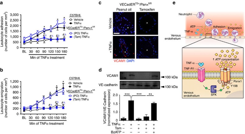
Inflammatory cell recruitment to local sites of tissue injury and/or infection is controlled by a plethora of signalling processes influencing cell-to-cell interactions between the vascular endothelial cells (ECs) in post-capillary venules and circulating leukocytes. Recently, ATP-sensitive P2Y purinergic receptors have emerged as downstream regulators of EC activation in vascular inflammation. However, the mechanism(s) regulating cellular ATP release in this response remains elusive. Here we report that the ATP-release channel Pannexin1 (Panx1) opens downstream of EC activation by TNF-α. This process involves activation of type-1 TNF receptors, recruitment of Src family kinases (SFK) and SFK-dependent phosphorylation of Panx1. Using an inducible, EC-specific Panx1 knockout mouse line, we report a previously unidentified role for Panx1 channels in promoting leukocyte adhesion and emigration through the venous wall during acute systemic inflammation, placing Panx1 channels at the centre of cytokine crosstalk with purinergic signalling in the endothelium.


Figures

Figure 1
Figure 1. Venous endothelial cells release ATP when activated by TNF-α.
(a) Schematic of ex vivo vascular perfusion assay. (b) TNF-α-induced ATP release from isolated murine mesenteric venules. TNF-α promoted a time- and dose-dependent increase in ATP release from the endothelium. *P<0.05, **P<0.01 and ***P<0.001 as compared with vehicle-perfused controls (n=4). (c) LDH release from isolated venules perfused with TNF-α or lysis buffer. (d) ATP release from isolated mesenteric venules (V) and paired arterioles (A) in response to TNF-α (50 ng ml−1) perfusion. ***P<0.001 versus venule (n=4). (e) Time course of ATP release from mesenteric venules following inhibition of TNFR1 with WP9QY (10 μM). *P<0.05 and **P<0.01 versus the corresponding vehicle time point (n=4). (f) Dose response of primary human venous (HUVEC and HSaVEC) and arterial (HAoEC and HCoAEC) ECs to TNF-α. HUVEC, human umbilical vein endothelial cell; HSaVEC, human saphenous vein endothelial cell; HAoEC, human aortic endothelial cell; HCoAEC, human coronary artery endothelial cell. *P<0.01 compared with unstimulated cells and #P<0.005 as compared with venous cells (n=5). (g) Time course of ATP release from cultured arterial and venous ECs. Cells were stimulated with 10 ng ml−1 TNF-α. (h) Dose response of HUVEC to TNF-α following inhibition of TNFR1 with WP9QY (10 μM). *P<0.05 as compared with vehicle control (n=5). All data are presented as mean±s.e.m. (error bars). Statistical analyses were performed using one-way analysis of variance.
Figure 2
Figure 2. Pannexin 1 channels mediate TNF-α-induced ATP release from venous ECs.
(a) Representative western blot of HUVEC treated with brefeldin A (BFA; 5 h) and subsequent cell surface biotinylation of membrane proteins. Plasma membrane localization of Panx1 and Cx43 were assessed using isoform-specific antibodies to each protein. (b) ATP release from BFA-treated HUVEC in response to TNF-α (10 ng ml−1) treatment for 30 min. (c,d) Time course of ATP release from HUVEC following inhibition of Panx1 channels with carbenoxolone (CBX: 50 μM) (c) and the Panx1 blocking peptide 10panx1 (200 μM) (d). *P<0.05 as compared with vehicle control (n=5). (e) Summary data of pharmacological inhibitors assessed for inhibition of TNF-α-induced ATP release from HUVEC. BFA (30 min): inhibition of vesicular release, Ruthenium red (RuR): antagonist of CALHM1 channels. Lanthanum (La3+): Cx hemichannel antagonist. *P<0.05 as compared with BFA, RuR and La3+ (n=5). (f) Representative western blots of siRNA knockdown of Panx1 and Cx43 in HUVEC. ***P<0.005 and ****P<0.001 versus control (n=3). (g) ATP release from Panx1 and Cx43 siRNA-treated HUVEC from f in response to TNF-α (10 ng ml−1). ***P<0.005 versus control (n=3). (h) Schematic representing the generation of an inducible, EC-specific Panx1 knockout mouse (VECadERT2+/Panx1fl/fl). (i) En face immunofluorescence micrographs of Panx1 (red) expression of endothelium from VECadERT2+/Panx1fl/fl mice injected with tamoxifen (Tam) or its vehicle peanut oil (PO) for 10 consecutive days. Nuclei are stained with 4,6-diamidino-2-phenylindole (DAPI) (blue). Scale bar, 10 μm. (j) Immuno-scanning electron micrographs (iSEM) of isolated mesenteric venules from VECadERT2+/Panx1fl/fl mice. Veins were immunolabelled for Panx1 (pseudo-coloured magenta) using an antibody against the extracellular region of the channel. Right panels are zoomed images of the left panels. Scale bar, 10 μm; enlarged boxes are 5 × 5 μm. (k) TNF-α-induced ATP release from isolated mesenteric venules from VECadERT2+/Panx1fl/fl mice injected with Tam or PO for 10 days. Venules were perfused with 50 ng ml−1 TNF-α or vehicle. *P<0.05 as compared with vehicle-perfused controls and #P<0.05 as compared with PO+TNF-α (n=4). All data are presented as mean±s.e.m. (error bars). Statistical analyses were performed using one-way analysis of variance. NS, not significant.
Figure 3
Figure 3. TNF-α induces Src family kinase-dependent activation of EC Panx1 channels.
(a) Western blot analysis of SFK activation in HUVEC in response to TNF-α stimulation (10 ng ml−1). A phospho-specific antibody against Y416 in SFKs (pY416SFK) was used as an indicator of SFK activation. SFK activation was blocked with the pharmacological antagonist PP2 (10 μM) but not by its inactive analogue PP3 (10 μM). Antibody specificity for the phosphorylated form of the kinases was confirmed by dephosphorylating proteins in cell lysates with alkaline phosphatase. *P<0.05 as compared with unstimulated control (lane 1) and #P<0.001 as compared with lane 1 (n=3). (b) TNF-α-induced ATP release from HUVEC following SFK inhibition with PP2. *P<0.05 versus control and PP3 treatments (n=5). (c) Topological schematic of Panx1 highlighting an epitope in the intracellular loop which contains tyrosine 198. This epitope was used to develop antibodies specific to the phosphorylated (pY198Panx1) and non-phosphorylated (Panx1-IL) forms of the protein. (d) Overlay of pY198Panx1 signal and Panx1-IL (total) signal as assessed by western blotting with LiCOR IRDye secondary antibodies. pY198Panx1 detects a single species at ∼55 kDa. (e) Western blot analysis of pY198Panx1 in HUVEC transfected with plasmids encoding c-Src and/or inhibitor of Src (i-Src). (f) Western blot analysis of Panx1 phosphorylation at Y198 in HUVECs stimulated with TNF-α (10 ng ml−1). Phospho-signal was normalized to total Panx1 expression using the Panx1-IL Ab. *P<0.05 compared with vehicle control (lane 1) and #P<0.01 compared with 5-min TNF-α stimulation (lane 4) (n=3). (g) TNF-α-induced ATP release from mesenteric venules treated with PP2 (10 μM) or PP3 (10 μM). *P<0.05 as compared with vehicle control (n=5). (h,i) Western blot analysis of TNF-α-induced SFK activation (h) and pY198Panx1 phosphorylation (i) in isolated mesenteric venules perfused with TNF-α (50 ng ml−1) for 30 min. (j) Immunofluorescence micrographs of pY198Panx1 in isolated mesenteric venule cross-sections. Venules were isolated from mice expressing endogenous Panx1 in the vascular wall (VECadERT2+/Panx1fl/fl+peanut oil) or mice with specific EC Panx1 deletion (VECadERT2+/Panx1fl/fl+tamoxifen) and stimulated with TNF-α. Asterisks indicate the vessel lumen and nuclei are stained with 4,6-diamidino-2-phenylindole (DAPI) (blue). Scale bar, 30 μm. All data are presented as mean±s.e.m. (error bars). Statistical analyses were performed using one-way analysis of variance.
Figure 4
Figure 4. EC Panx1 channels promote leukocyte adhesion and emigration.
(a,b) Quantification of endogenous leukocyte adhesion (a) and emigration (b) in wild-type (WT) C57Bl/6J mice and mice lacking Panx1 specifically in the endothelium (VECadERT2+/Panx1fl/fl) in the acute inflammatory response. Acute inflammation was induced by topically applying recombinant murine TNF-α to exteriorized cremaster muscles in anaesthetized mice. *P<0.01 as compared with baseline, #P<0.005 as compared with C57Bl/6J mice treated with TNF-α and ^P<0.01 as compared with PO-injected VECadERT2+/Panx1fl/fl mice treated with TNF-α by two-way analysis of variance (ANOVA) (n=5–6 mice per group). (c) Immunofluorescence micrographs for VCAM1 expression in isolated mesenteric venules from VECadERT2+/Panx1fl/fl mice (PO or Tam injected) treated with vehicle or TNF-α (50 ng ml−1) for 2 h. * indicates the vessel lumen and nuclei are stained with 4,6-diamidino-2-phenylindole (DAPI) (blue). Scale bar, 30 μm. (d) Western blot analysis of VCAM1 expression in isolated mesenteric venules from VECadERT2+/Panx1fl/fl mice following TNF-α (50 ng ml−1) treatment. Exogenous BzATP (10 μM) was applied to assess the potential to rescue VCAM1 upregulation. **P<0.01 and ***P<0.005 by one-way ANOVA (n=3). (e) Mechanism of TNF-α-induced ATP release from venous ECs in the acute inflammatory response. All data are presented as mean±s.e.m. (error bars).

Similar articles

Cited by

References

    1. Ley K., Laudanna C., Cybulsky M. I. & Nourshargh S. Getting to the site of inflammation: the leukocyte adhesion cascade updated. Nat. Rev. Immunol. 7, 678–689 (2007). - PubMed
    1. Ralevic V. & Burnstock G. Receptors for purines and pyrimidines. Pharmacol. Rev. 50, 413–492 (1998). - PubMed
    1. Zerr M. et al. Major contribution of the P2Y(1)receptor in purinergic regulation of TNFalpha-induced vascular inflammation. Circulation 123, 2404–2413 (2011). - PubMed
    1. Riegel A. K. et al. Selective induction of endothelial P2Y6 nucleotide receptor promotes vascular inflammation. Blood 117, 2548–2555 (2011). - PMC - PubMed
    1. Hyman M. C. et al. Self-regulation of inflammatory cell trafficking in mice by the leukocyte surface apyrase CD39. J. Clin. Invest. 119, 1136–1149 (2009). - PMC - PubMed

Publication types

MeSH terms