Review
. 2015 Aug;3(8):827-35.
doi: 10.1158/2326-6066.CIR-15-0143.

Reflections on the Histopathology of Tumor-Infiltrating Lymphocytes in Melanoma and the Host Immune Response

Affiliations
Review

Reflections on the Histopathology of Tumor-Infiltrating Lymphocytes in Melanoma and the Host Immune Response

Martin C Mihm Jr et al. Cancer Immunol Res. 2015 Aug.

Abstract

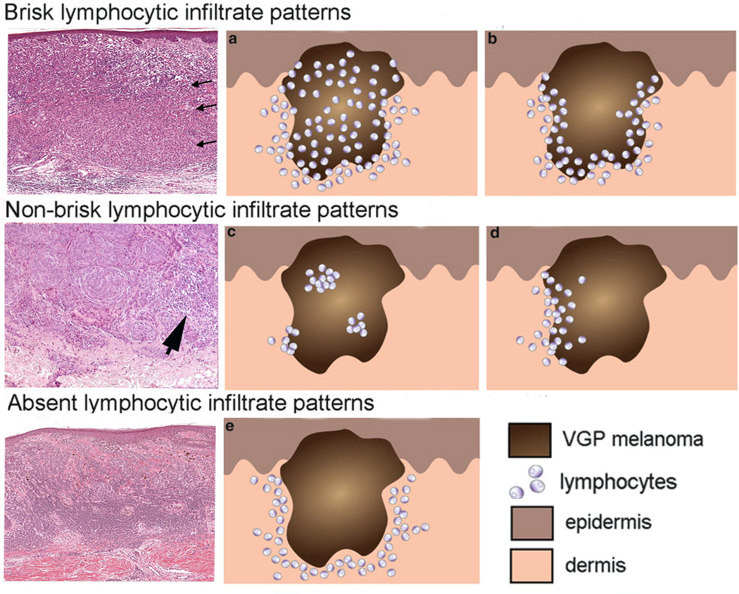
In the past five decades, the role for lymphocytes in host immune response to tumors has been shown, at least in some patients, to be a critical component in disease prognosis. Also, the heterogeneity of lymphocytes has been documented, including the existence of regulatory T cells that suppress the immune response. As the functions of lymphocytes have become better defined in terms of antitumor immunity, specific targets on lymphocytes have been uncovered. The appreciation of the role of immune checkpoints has also led to therapeutic approaches that illustrate the effectiveness of blocking negative regulators of the antitumor immune response. In this Masters of Immunology article, we trace the evolution of our understanding of tumor-infiltrating lymphocytes and discuss their role in melanoma prognosis from the very basic observation of their existence to the latest manipulation of their functions with the result of improvement of the host response against the tumor.


Figures

Figure 1
Figure 1
Melanoma infiltrated with various immune cell types. Panel A: Superficial spreading melanoma with multiple lymphocytes (arrow L) infiltrate amidst melanoma cells (arrow) in papillary dermis (Hematoxylin and eosin-stained light micrographs; 40x.) Panel B: Dense melanophages (MP) with scattered lymphocytes (blue) and fibrosis represent area of regression flanked by the tumor (magenta). Panel C: Nodule of metastatic melanoma with dense lymphoid infiltrate (L arrowhead). Note the striking interaction of lymphocytes with melanoma cells, and areas of melanoma cell nuclear pyknosis.
Figure 1
Figure 1
Melanoma infiltrated with various immune cell types. Panel A: Superficial spreading melanoma with multiple lymphocytes (arrow L) infiltrate amidst melanoma cells (arrow) in papillary dermis (Hematoxylin and eosin-stained light micrographs; 40x.) Panel B: Dense melanophages (MP) with scattered lymphocytes (blue) and fibrosis represent area of regression flanked by the tumor (magenta). Panel C: Nodule of metastatic melanoma with dense lymphoid infiltrate (L arrowhead). Note the striking interaction of lymphocytes with melanoma cells, and areas of melanoma cell nuclear pyknosis.
Figure 2
Figure 2
Vertical growth phase melanoma with striking lymphocyte (yellow L arrows)-melanoma cell satellitosis with evidence of degeneration of affected melanoma cells (arrow M).
Figure 3
Figure 3
Brisk, non-brisk, and absent lymphocytic infiltration in primary melanoma nodules. The three photomicrographs in the left column exhibit the brisk, non-brisk and absent patterns. The brisk pattern is defined by lymphocytes present throughout the tumor as shown by arrows in the top left panel. Non-brisk is defined by scattered, focal groups of lymphocytes (arrow). Panel 3: Absent is defined by no lymphocyte infiltration in the tumor. Panels (a, b, c, d, e) in the middle and right columns are schematic renderings of different types of TIL patterns in primary melanoma. The brisk lymphocytic response may involve the entire tumor nodule (panel a) or be present along the advancing edge of the entire peripheral nodule (panel b). The focal nature of the non-brisk pattern is demonstrated in panel c and d. In the rendering of the absent lymphocytic response, there are either no lymphocytes in the tumor, or if lymphocytes are present (panel e) they do not interact with melanoma cells. Panels a, b, c, d, e are adapted from figure 4 of chapter 16 by Schatton and colleagues, entitled “Tumor Infiltrating Lymphocytes and their Significance in Melanoma Prognosis,” in Molecular Diagnostics for Melanoma. Thurin M, Marincola FM eds; Humana Press. 2014, 287–324.
Figure 4
Figure 4
The image depicts in light microscopy the Ki-67 immunohistochemical staining of a section of a typical TL-ELN (center) within a stage IV (non-locoregional), visceral melanoma metastasis. The tissue was stained using the avidin-biotin complex method with retrieval under high pH. Prediluted antibody to Ki-67 [Ventana Medical System, Inc.; anti-Ki-67 (–9) primary antibody is a rabbit monoclonal antibody (IgG) directed against C-terminal portion of Ki-67 antigen.] was used for the proliferative analysis of the TL-ELN. Lymphocytic proliferation within and surrounding the follicle (brown staining cells) was found to contain both B and T lymphocytes, suggesting newly formed and/or activated TL-ELNs within the tumor microenvironment. (Image was generated by Dr. Jane Messina of the Departments of Anatomic Pathology and Cutaneous Oncology at the Moffitt Cancer Center.

References

    1. Virchow R. Die Krankhaften Geschwulste: Dressig Vorlesungen, gehalten waehrend des Wintersemesters 1862–1863 an der Universitaet zu Berlin: Vorlesungen ueber Pathologie: Verlag von August Hirschwald. 1863.
    1. Ehrlich P. Experimentelle studien an maestumoren. Zeitschrift f Krebsforschung. 1907;5:59–81.
    1. Coley W. The treatment of malignant tumors by repeated inoculations of Erysipelas, with a report of ten original cases. Am J Med SCi. 1989;105:487–511. - PubMed
    1. Moore OS, Jr, Foote FW., Jr The relatively favorable prognosis of medullary carcinoma of the breast. Cancer. 1949;2:635–642. - PubMed
    1. Brownstein S, Sheikh KM, Lewis MG. Immunological studies in patients with malignant melanoma of the uvea. Can J Ophthalmol. 1977;12:16–23. - PubMed

Publication types

MeSH terms