

Rate of Contrast Extravasation on Computed Tomographic Angiography Predicts Hematoma Expansion and Mortality in Primary Intracerebral Hemorrhage
- PMID: 26243220
- PMCID: PMC4550492
- DOI: 10.1161/STROKEAHA.115.009659
Rate of Contrast Extravasation on Computed Tomographic Angiography Predicts Hematoma Expansion and Mortality in Primary Intracerebral Hemorrhage
Abstract
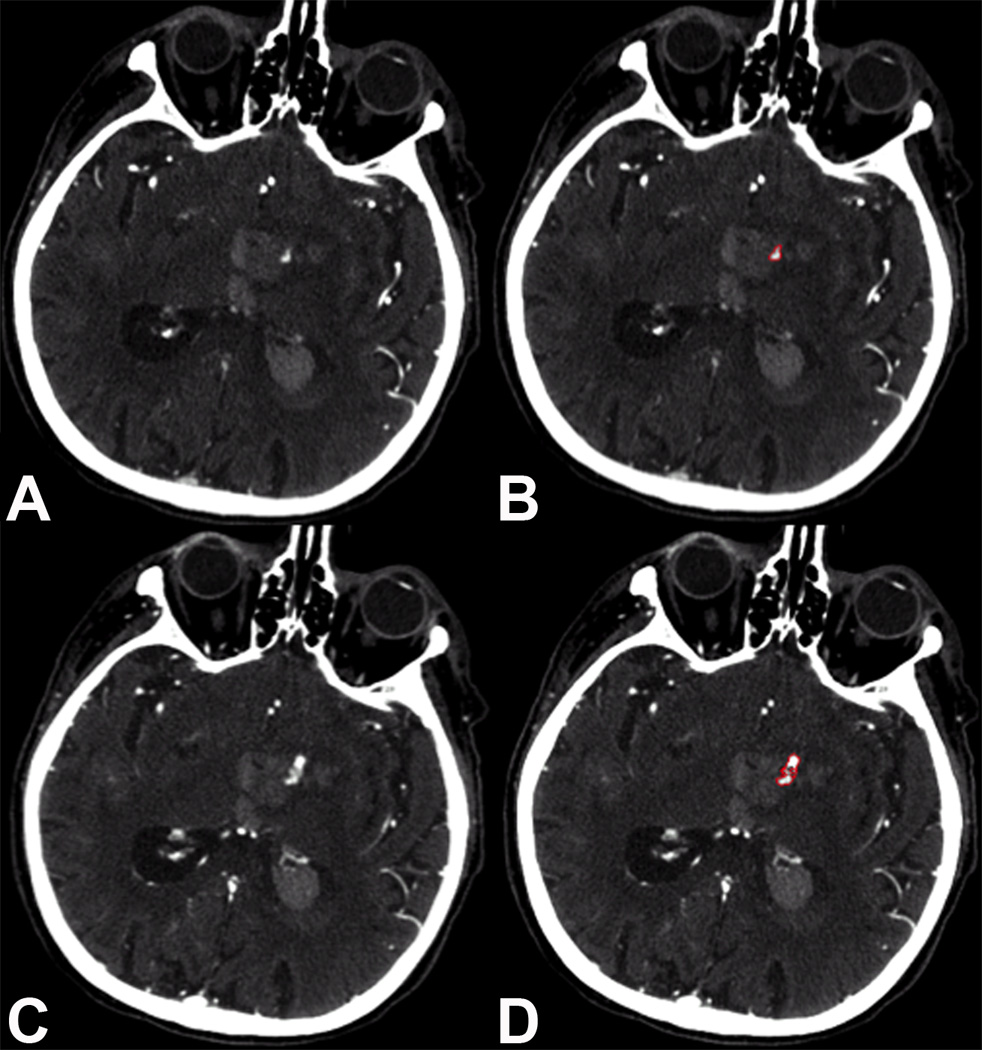
Background and purpose: In primary intracerebral hemorrhage, the presence of contrast extravasation after computed tomographic angiography (CTA), termed the spot sign, predicts hematoma expansion and mortality. Because the biological underpinnings of the spot sign are not fully understood, we investigated whether the rate of contrast extravasation, which may reflect the rate of bleeding, predicts expansion and mortality beyond the simple presence of the spot sign.
Methods: Consecutive intracerebral hemorrhage patients with first-pass CTA followed by a 90-second delayed postcontrast CT (delayed CTA) were included. CTAs were reviewed for spot sign presence by 2 blinded readers. Spot sign volumes on first-pass and delayed CTA and intracerebral hemorrhage volumes were measured using semiautomated software. Extravasation rates were calculated and tested for association with hematoma expansion and mortality using uni- and multivariable logistic regressions.
Results: One hundred and sixty-two patients were included, 48 (30%) of whom had ≥1 spot sign. Median spot sign volume was 0.04 mL on first-pass CTA and 0.4 mL on delayed CTA. Median extravasation rate was 0.23 mL/min overall and 0.30 mL/min among expanders versus 0.07 mL/min in nonexpanders. Extravasation rates were also significantly higher in patients who died in hospital: 0.27 mL/min versus 0.04 mL/min. In multivariable analysis, the extravasation rate was independently associated with in-hospital mortality (odds ratio, 1.09 [95% confidence interval, 1.04-1.18], P=0.004), 90-day mortality (odds ratio, 1.15 [95% confidence interval, 1.08-1.27]; P=0.0004), and hematoma expansion (odds ratio, 1.03 [95% confidence interval, 1.01-1.08]; P=0.047).
Conclusions: Contrast extravasation rate, or spot sign growth, further refines the ability to predict hematoma expansion and mortality. Our results support the hypothesis that the spot sign directly measures active bleeding in acute intracerebral hemorrhage.
Keywords: CT angiography; cerebral hemorrhage; intracerebral hemorrhage; mortality.
© 2015 American Heart Association, Inc.
Figures

References
-
- van Asch CJ, Luitse MJ, Rinkel GJ, van der Tweel I, Algra A, Klijn CJ. Incidence, case fatality, and functional outcome of intracerebral haemorrhage over time, according to age, sex, and ethnic origin: a systematic review and meta-analysis. Lancet Neurol. 2010;9:167–176. - PubMed
-
- Broderick JP, Brott TG, Duldner JE, Tomsick T, Huster G. Volume of intracerebral hemorrhage. A powerful and easy-to-use predictor of 30-day mortality. Stroke. 1993;24:987–993. - PubMed
-
- Davis SM, Broderick J, Hennerici M, Brun NC, Diringer MN, Mayer SA, et al. Hematoma growth is a determinant of mortality and poor outcome after intracerebral hemorrhage. Neurology. 2006;66:1175–1181. - PubMed
-
- Delcourt C, Huang Y, Arima H, Chalmers J, Davis SM, Heeley EL, et al. Hematoma growth and outcomes in intracerebral hemorrhage: the INTERACT1 study. Neurology. 2012;79:314–319. - PubMed
Publication types
MeSH terms
Grants and funding
LinkOut - more resources
Full Text Sources
