Observational Study
. 2015 Oct;56(10):2010-8.
doi: 10.1194/jlr.M060970. Epub 2015 Aug 4.

Protective associations of HDL with blood-brain barrier injury in multiple sclerosis patients

Affiliations
Observational Study

Protective associations of HDL with blood-brain barrier injury in multiple sclerosis patients

Kelly Fellows et al. J Lipid Res. 2015 Oct.

Abstract

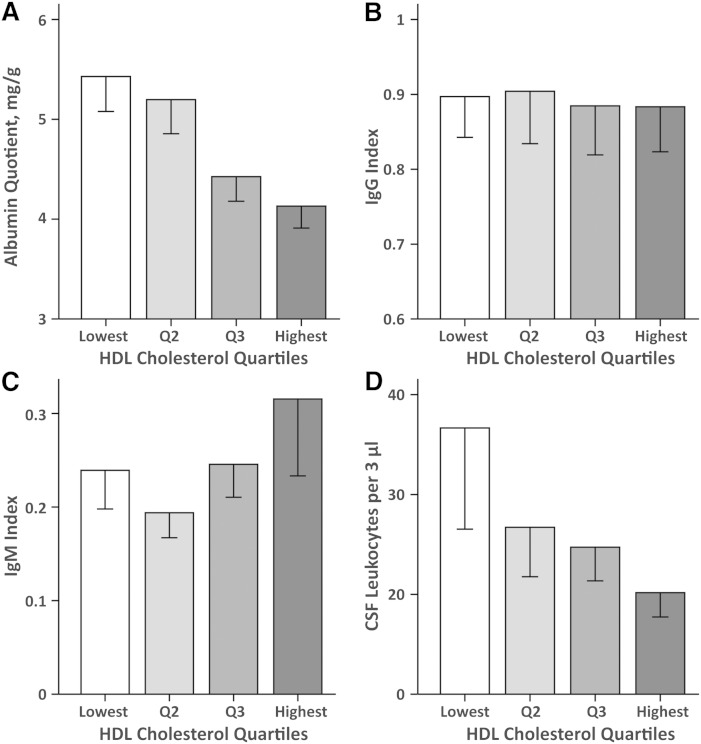
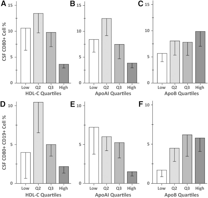
The purpose of this work was to investigate the associations of serum cholesterol and apolipoproteins with measures of blood-brain barrier (BBB) permeability and CNS inflammation following the first clinical demyelinating event. This study included 154 patients [67% female; age, 29.5 ± 8.2 years (mean ± SD)] enrolled in a multi-center study of interferon β1-a treatment following the first demyelinating event. Blood and cerebrospinal fluid (CSF) were obtained at screening prior to treatment. A comprehensive serum lipid profile and multiple surrogate markers of BBB breakdown and CNS immune activity were obtained. Higher levels of serum HDL cholesterol (HDL-C) and ApoA-I were associated with lower CSF total protein level, CSF albumin level, albumin quotient, and CSF IgG level (all P ≤ 0.001 for HDL-C and all P < 0.01 for ApoA-I). HDL-C was also associated with CSF CD80+ (P < 0.001) and with CSF CD80+CD19+ (P = 0.007) cell frequencies. Higher serum HDL is associated with lower levels of BBB injury and decreased CD80+ and CD80+CD19+ cell extravasation into the CSF. HDL may potentially inhibit the initiation and/or maintenance of pathogenic BBB injury following the first demyelinating event.

Keywords: apolipoproteins; cholesterol; clinically isolated syndrome; high density lipoprotein.


Figures

Fig. 1.
Fig. 1.
Associations of HDL-C quartiles with albumin quotient (A), IgG index (B), IgM index (C), and CSF leukocytes (D). The quartile boundaries were: the lowest quartile corresponds to HDL-C ≤ 56.27 mg/dl, 56.27 mg/dl < quartile 2 (Q2) ≤ 66.25 mg/dl, 66.25 mg/dl < quartile 3 (Q3) ≤ 80.69 mg/dl, and the highest quartile > 80.69 mg/dl. The bars compare mean values of the CSF measures shown on the y axis for the quartiles of HDL-C shown on the x axis. The error bars indicate the standard error of the mean.
Fig. 2.
Fig. 2.
Associations of ApoA-I quartiles with albumin quotient (A), IgG index (B), IgM index (C), and CSF leukocytes (D). The ApoA-I quartile boundaries were: the lowest quartile corresponds to ApoA-I ≤ 128.3 mg/dl, 128.3 mg/dl < quartile 2 (Q2) ≤ 151.1 mg/dl, 151.1 mg/dl < quartile 3 (Q3) ≤ 175.8 mg/dl, and the highest quartile > 175.8 mg/dl. The bars compare mean values of the CSF measures shown on the y axis to the quartiles of ApoA-I shown on the x axis. The error bars indicate the standard error of the mean.
Fig. 3.
Fig. 3.
Associations of mean CSF CD80+ (A–C) and CSF CD80+CD19+ (D–F) cell frequencies in percent with HDL-C quartiles, ApoA-I quartiles, and ApoB quartiles. A, D: The HDL-C quartile boundaries were: the lowest quartile corresponds to HDL-C ≤ 56.27 mg/dl, 56.27 mg/dl < quartile 2 (Q2) ≤ 66.25 mg/dl, 66.25 mg/dl < quartile 3 (Q3) ≤ 80.69 mg/dl, and the highest quartile > 80.69 mg/dl. B, E: The ApoA-I quartile boundaries were: the lowest quartile corresponds to ApoA-I ≤ 128.3 mg/dl, 128.3 mg/dl < quartile 2 ≤ 151.1 mg/dl, 151.1 mg/dl < quartile 3 ≤ 175.8 mg/dl, and the highest quartile > 175.8 mg/dl. C, F: The ApoB quartile boundaries were: the lowest quartile corresponds to ApoB ≤ 54.0 mg/dl, 54.0 mg/dl < quartile 2 ≤ 70.75 mg/dl, 70.75 mg/dl < quartile 3 ≤ 87.0 mg/dl, and the highest quartile > 87.0 mg/dl. The bars compare mean values of the CSF cell frequencies in percent shown on the y axis to the quartiles of HDL-C, ApoA-I, or ApoB on the x axis. The error bars indicate the standard error of the mean.
Fig. 4.
Fig. 4.
Associations of albumin quotient (A, B) and CSF leukocytes (C, D) with CEL number and T2-LV MRI measures. The bars compare mean values of the MRI measures shown on the y axis for the quartiles of the CSF variable shown on the x axis. A, B: The albumin quotient quartile boundaries were: the lowest quartile corresponds to albumin quotient ≤ 3.36 mg/g, 3.36 mg/g < quartile 2 (Q2) ≤ 4.25 mg/g, 4.25 mg/g < quartile 3 (Q3) ≤ 5.58 mg/g, and the highest quartile > 5.58 mg/g. C, D: The CSF leukocytes quartile boundaries were: the lowest quartile corresponds to CSF leukocytes ≤ 9.0/3 μl, 10.0/3 μl ≤ quartile 2 ≤ 17.0/3 μl, 18.0/3 μl ≤ quartile 3 ≤ 33.5/3 μl, and the highest quartile > 33.5/3 μl. The error bars indicate the standard error of the mean.

References

    1. Minagar A., Alexander J. S. 2003. Blood-brain barrier disruption in multiple sclerosis. Mult. Scler. 9: 540–549. - PubMed
    1. Ortiz G. G., Pacheco-Moises F. P., Macias-Islas M. A., Flores-Alvarado L. J., Mireles-Ramirez M. A., Gonzalez-Renovato E. D., Hernandez-Navarro V. E., Sanchez-Lopez A. L., Alatorre-Jimenez M. A. 2014. Role of the blood-brain barrier in multiple sclerosis. Arch. Med. Res. 45: 687–697. - PubMed
    1. Lucchinetti C. F., Popescu B. F., Bunyan R. F., Moll N. M., Roemer S. F., Lassmann H., Bruck W., Parisi J. E., Scheithauer B. W., Giannini C., et al. 2011. Inflammatory cortical demyelination in early multiple sclerosis. N. Engl. J. Med. 365: 2188–2197. - PMC - PubMed
    1. Werring D. J., Brassat D., Droogan A. G., Clark C. A., Symms M. R., Barker G. J., MacManus D. G., Thompson A. J., Miller D. H. 2000. The pathogenesis of lesions and normal-appearing white matter changes in multiple sclerosis: a serial diffusion MRI study. Brain. 123: 1667–1676. - PubMed
    1. Holmøy T. 2009. The discovery of oligoclonal bands: a 50-year anniversary. Eur. Neurol. 62: 311–315. - PubMed

Publication types

MeSH terms