. 2015 Oct;169(2):1179-91.
doi: 10.1104/pp.15.00229. Epub 2015 Aug 4.

Rice TUTOU1 Encodes a Suppressor of cAMP Receptor-Like Protein That Is Important for Actin Organization and Panicle Development

Affiliations

Rice TUTOU1 Encodes a Suppressor of cAMP Receptor-Like Protein That Is Important for Actin Organization and Panicle Development

Jiaoteng Bai et al. Plant Physiol. 2015 Oct.

Abstract

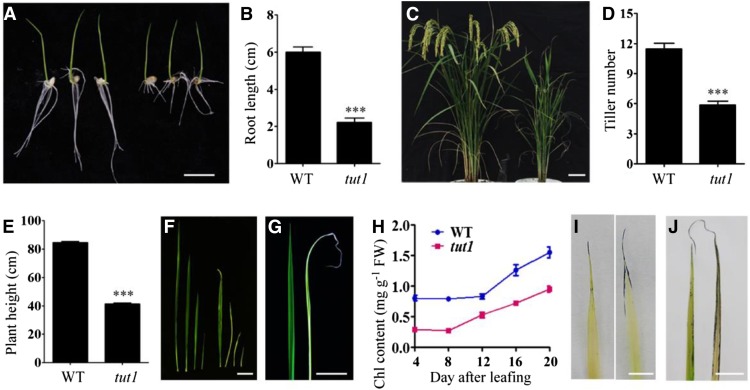
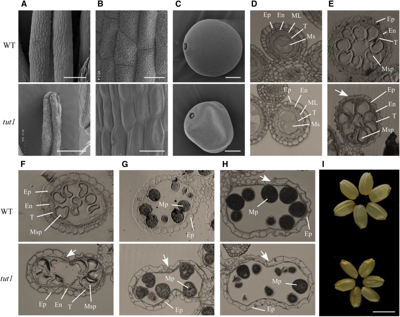
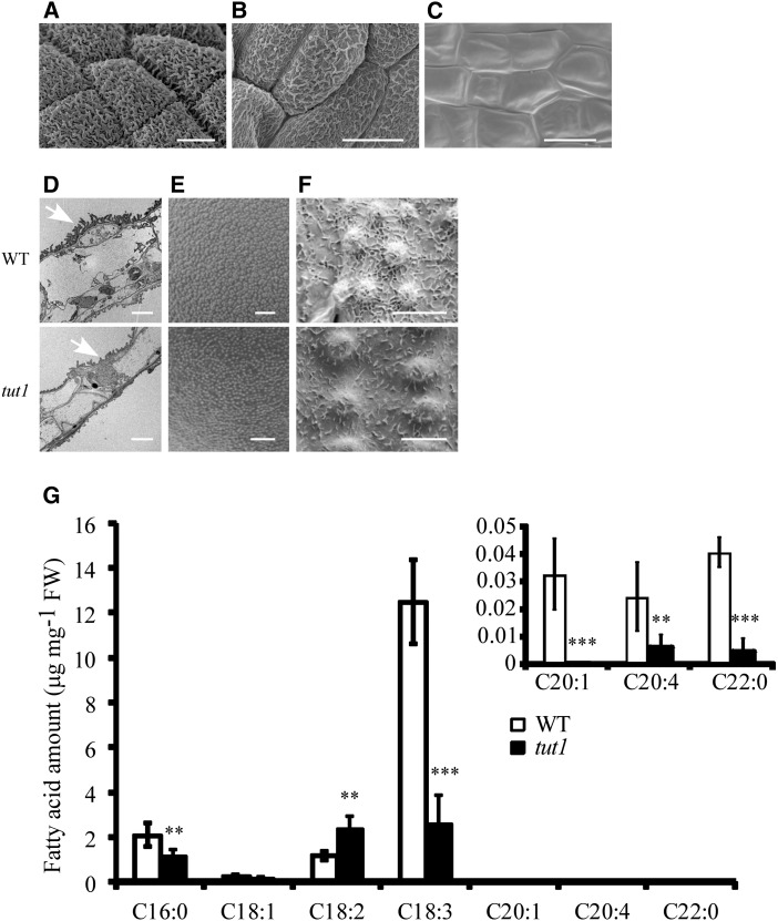
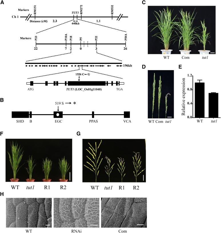
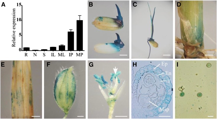
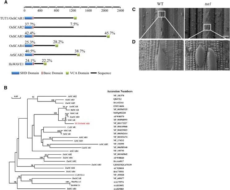
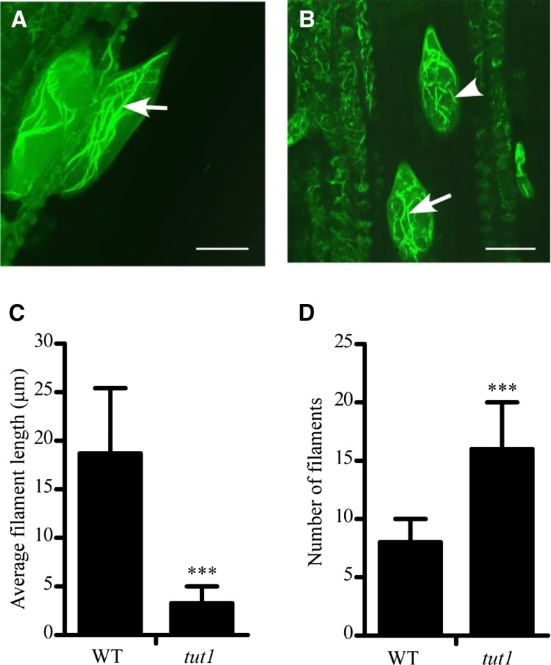
Panicle development, a key event in rice (Oryza sativa) reproduction and a critical determinant of grain yield, forms a branched structure containing multiple spikelets. Genetic and environmental factors can perturb panicle development, causing panicles to degenerate and producing characteristic whitish, small spikelets with severely reduced fertility and yield; however, little is known about the molecular basis of the formation of degenerating panicles in rice. Here, we report the identification and characterization of the rice panicle degenerative mutant tutou1 (tut1), which shows severe defects in panicle development. The tut1 also shows a pleiotropic phenotype, characterized by short roots, reduced plant height, and abnormal development of anthers and pollen grains. Molecular genetic studies revealed that TUT1 encodes a suppressor of cAMP receptor/Wiskott-Aldrich syndrome protein family verprolin-homologous (SCAR/WAVE)-like protein. We found that TUT1 contains conserved functional domains found in eukaryotic SCAR/WAVE proteins, and was able to activate Actin-related protein2/3 to promote actin nucleation and polymerization in vitro. Consistently, tut1 mutants show defects in the arrangement of actin filaments in trichome. These results indicate that TUT1 is a functional SCAR/WAVE protein and plays an important role in panicle development.


Figures

Figure 1.
Figure 1.
Phenotypes of tut1. A, Seedling phenotype of the wild type (cv Zhonghua 11, left three seedlings) and tut1 (right three seedlings). Seeds were germinated at 30°C for 3 d and then transferred to a culture chamber with a 16-h/8-h photoperiod at 30°C. Bar = 1 cm. B, Statistical analysis of root length in the wild type (WT) and tut1 as shown in A. The data presented are mean values of two independent experiments (n > 45). Error bars indicate sd; ***, P < 0.01 (Student’s t test). C, Phenotypes of wild-type (left) and tut1 (right) mutants at the grain-filling stage. Bar = 5 cm. D and E, Statistical analysis of tiller number and plant height at maturity of the wild type and tut1. The data presented are mean values (n > 50). Error bars indicate sd; ***, P < 0.01 (Student’s t test). F, Leaves of the wild type (left four leaves) and tut1 (right four leaves) at different growth stages. Bar = 5 cm. G, Leaves of the wild type (left) and tut1 (right) at 10 d after leafing. Bar = 2 cm. H, Analysis of chlorophyll (Chl) content of wild-type and tut1 leaves at different times after leafing. Blue line indicates the wild type, red line indicates tut1. Leaves at 4, 8, 12, 16, and 20 d after leafing were used in the analysis. Fresh leaves were used for measurement of chlorophyll contents. Two technical repeats were made in the test. Error bars indicate sd. I, Cell death analysis of wild-type (left) and tut1 (right) seedling leaves using Evans blue staining. Bar = 0.5 cm. J, ROS observation of wild-type (left) and tut1 (right) mature leaves using nitroblue tetrazolium (NBT) staining. FW, Fresh weight. Bar = 2 cm.
Figure 2.
Figure 2.
Morphology of panicle and spikelet in tut1. A, Panicle rachis of wild-type (WT; left) and tut1 (right) plants at the grain-filling stage. Bar = 2 cm. B, The expanding panicle branch of wild-type (left) and tut1 (right) plants at the grain-filling stage. The part of the tut1 panicle shown in the white box was enlarged. The arrows show the degenerated branches and spikelets in the tut1. Bars = 2 cm. C, ROS analysis of the wild type (bottom) and tut1 (top) by NBT staining. Bar = 2 mm. D, ROS analysis of spikelets showing different degrees of degeneration in tut1 at the heading stage by NBT staining. Bar = 2 mm. E, Comparison of wild-type (left one) and tut1 spikelets (right three) after removing the lemma and palea at the heading stage. Right three spikelets, named Type 1, Type 2 and Type 3, show varying degrees of developmental defects in tut1. Bar = 1 mm. F to K, Scanning electron microscopy (SEM) observation of spikelet at the tip of the panicle at stage In7 (F), stage In8 (arrows indicate the apical spikelets in the wild type and the tut1 [G]), stages In8 and In9 (H; the white square frame region shown in H is enlarged in I), and stage In9 (J; the white square frame region shown in J is enlarged in K). Bars = 100 μm (F and K), 400 μm (G), 200 μm (H), 10 μm (I), and 500 μm (J).
Figure 3.
Figure 3.
SEM and semithin section analysis of wild-type and tut1 anther morphology. A to C, SEM observation of mature anthers of the wild type (WT) and tut1 (A), enlarged view of the anther surface of the wild type and tut1 (B), and mature pollen grain of the wild type and the tut1 (C). Bars = 200 μm (A), 20 μm (B), and 10 μm (C). D to H, The cross sections were stained with toluidine blue. Ep, Epidermis; En, endothecium; ML, middle layer; Ms, microsporocyte; Msp, microspore; Mp, mature pollen; T, tapetum. Cross section of single locule at the microspore mother cell stage; Ep, En, ML, T, and Ms are indicated (D); vacuolated pollen stage; Ep, En, T, and Msp are indicated (E); pollen first mitosis stage; Ep, En, T, and Msp are indicated (F); pollen second mitosis stage; Ep and Mp are indicated (G); and mature pollen stage; Ep and Mp are indicated (H). The arrows show the degenerated epidermal cell. I, Phenotype of mature seeds without glumes of the wild type (top) and tut1 (bottom). Bar = 5 mm.
Figure 4.
Figure 4.
Cuticle formation and fatty acid metabolism in the wild type (WT) and tut1. A to C, SEM observation of mature anther cutin of the wild type (A) and tut1 at the middle panicle (B) and apical panicle (C). tut1 anthers showing different degrees of degeneration are shown in B and C. Bars = 10 μm. D, Transmission electron microscopy observation of wild-type and tut1 mature anthers. Arrowhead indicates cuticle. Bar = 5 μm. E and F, SEM observation of the surface of mature pollen grains (E) and the cuticle of mature leaves (F) of the wild type and tut1. Bars = 2 μm (E) and 5 μm (F). G, Analysis of fatty acid contents in the wild type and tut1 using gas chromatography. Fresh panicles at the heading stage of the wild type and tut1 were used in the measurement. C17 was added as an internal standard. Seven types of fatty acid, C16:0, C18:1, C18:2, C18:3, C20:1, C20:4, and C22:0, were measured, and three biological repeats were conducted. FW, Fresh weight. Error bars indicate sd. **, P < 0.05; ***, P < 0.01 (Student’s t test).
Figure 5.
Figure 5.
Map-based cloning of TUT1. A, TUT1 maps on chromosome 1. TUT1 was roughly mapped to a 440-kb interval between the SSR markers RM151 and RM272. The number of recombinants between the molecular markers and TUT1 are indicated. TUT1 was fine mapped to an interval of about 196 kb between the single nucleotide polymorphism (SNP) markers P18 and P22. This region contained 28 predicted genes. A one-nucleotide transversion mutation was found in a gene annotated as a SCAR-like protein2 (LOC_Os01g11040). The mutation site at nucleotide 1,556 (C–G) in the sixth exon is indicated by the asterisk. Ch 1, Chromosome 1. B, Conserved domain organization of TUT1, showing the SCAR/WAVE homolog domain (SHD), basic domain (B), envelope glycoprotein C (EGC) domain, pantetheine attachment site (PPAS), and VCA domain as black rectangles. The mutation alters the 519th codon (Ser) to a termination codon (shown as a white dot in the EGC domain). The asterisk indicates the termination codon. C, Phenotypes of the wild type (WT), complemented transgenic line (Com), and tut1 at the heading stage. Bar = 10 cm. D, Mature panicle phenotypes of the wild type, complemented line, and tut1. Bar = 2 cm. E, TUT1 expression analysis of the wild type and tut1 by quantitative reverse transcription (qRT)-PCR. F, Morphology of wild type, tut1, and RNA interference (RNAi) transgenic lines (R1 and R2) at the heading stage. Bar = 20 cm. G, Panicles of wild type, tut1, and RNAi transgenic lines (R1 and R2) at the flowering stage. Bar = 3 cm. H, Anther cuticle of wild type, tut1, and representative complemented line. Bars = 20 cm (F), 3 cm (G), and 5 μm (H).
Figure 6.
Figure 6.
Spatial expression pattern of TUT1. A, TUT1 mRNA levels were measured by qRT-PCR. Total RNA was extracted from the root (R), node (N), stem (S), immature leaf (IL), mature leaf (ML), immature panicle (IP), and mature panicle (MP) of the wild type. B to I, Expression of TUT1, as revealed by promoter-GUS fusion analysis in transgenic plants. GUS activity was observed in the mature glume, seed, and seed germ (B), root, coleoptile, and seedling (C); basal node of the shoot (D), leaf (E), and glume at the heading stage (F); and anther filaments and anthers. The arrow indicates the pistil (G). GUS staining of a cross section of the anther, with epidermis (Ep), endothecium (En), tapetum (T), and microspore (Msp) indicated (H) and mature pollen (I). Bars = 2 mm (B, D, and E), 4 mm (C), 1 mm (F and G), and 30 μm (H and I).
Figure 7.
Figure 7.
TUT1 belongs to the SCAR/WAVE family. A, Domain structure of SCAR/WAVE proteins. The rice genome encodes four SCAR proteins: TUT1/OsSCAR1, OsSCAR2, OsSCAR3, and OsSCAR4. All four have SHD and basic domains, but OsSCAR2 lacks the VCA domain. Percentages above each domain indicate the amino acid sequence identities of the SHD and VCA domains in each protein compared with TUT1. The domains are color coded: SHD (blue), basic domain (red), VCA domain (green), and the black line represents the amino acid sequence. B, A Phylogenetic tree of TUT1 and its homologous proteins in different species. The phylogenetic tree was constructed using DNAMAN software with 1,000 bootstrapping trials. Bootstrapping values over 50% and the scale bar are shown in the phylogenetic tree. The tree was constructed using the distance method with maximum likelihood. TUT1 is shown in red. Accession numbers for each sequence are shown. Species are as indicated: A. tauschii (Aet), Arabidopsis (At), B. distachyon (Bd), Caenorhabditis elegans (Ce), Cucumis sativus (Cs), Dictyostelium discoideum (Dd), Drosophila melanogaster (Dm), Glycine max (Gm), Homo sapiens (Hs), Mus musculus (Mm), rice (Os), S. italica (Si), Solanum lycopersicum (Sl), Solanum tuberosum (St), S. bicolor (Sb), and maize (Zm). C and D, Trichome phenotype of the wild type (WT) and tut1 shown by SEM. Mature trichomes of the wild type and tut1 are shown in C. Enlarged view of wild-type trichome and tut1 trichome (D) from regions indicated by white squares in C. Bars = 100 μm (C) and 10 μm (D).
Figure 8.
Figure 8.
F-actin organization in the wild type (WT) and tut1. A and B, F-actin organization in wild-type (A) and tut1 (B) mutant leaf trichomes visualized by staining with Alexa Fluor 488-phalloidin. Arrow indicates long actin filaments and arrowhead indicates fragmented F-actin. Bars = 20 μm. C, Analysis of average filament length in the wild type and tut1. ***, P < 0.01 (Student’s t test). D, Analysis of number of filaments in trichomes of the wild type and tut1. ***, P < 0.01 (Student’s t test). Over 20 trichomes were used in these analyses.

References

    1. Ashikari M, Sakakibara H, Lin S, Yamamoto T, Takashi T, Nishimura A, Angeles ER, Qian Q, Kitano H, Matsuoka M (2005) Cytokinin oxidase regulates rice grain production. Science 309: 741–745 - PubMed
    1. Basu D, Le J, El-Essal SD, Huang S, Zhang C, Mallery EL, Koliantz G, Staiger CJ, Szymanski DB (2005) DISTORTED3/SCAR2 is a putative Arabidopsis WAVE complex subunit that activates the Arp2/3 complex and is required for epidermal morphogenesis. Plant Cell 17: 502–524 - PMC - PubMed
    1. Beltzner CC, Pollard TD (2008) Pathway of actin filament branch formation by Arp2/3 complex. J Biol Chem 283: 7135–7144 - PubMed
    1. Bird D, Beisson F, Brigham A, Shin J, Greer S, Jetter R, Kunst L, Wu X, Yephremov A, Samuels L (2007) Characterization of Arabidopsis ABCG11/WBC11, an ATP binding cassette (ABC) transporter that is required for cuticular lipid secretion. Plant J 52: 485–498 - PubMed
    1. Blanchoin L, Amann KJ, Higgs HN, Marchand JB, Kaiser DA, Pollard TD (2000) Direct observation of dendritic actin filament networks nucleated by Arp2/3 complex and WASP/Scar proteins. Nature 404: 1007–1011 - PubMed

Publication types

MeSH terms

LinkOut - more resources