. 2016 Jan;31(1):69-81.
doi: 10.1093/mutage/gev055. Epub 2015 Aug 4.

Identification of genotoxic compounds using isogenic DNA repair deficient DT40 cell lines on a quantitative high throughput screening platform

Affiliations

Identification of genotoxic compounds using isogenic DNA repair deficient DT40 cell lines on a quantitative high throughput screening platform

Kana Nishihara et al. Mutagenesis. 2016 Jan.

Abstract

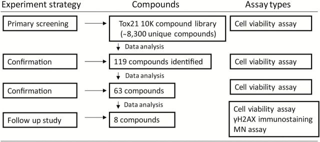
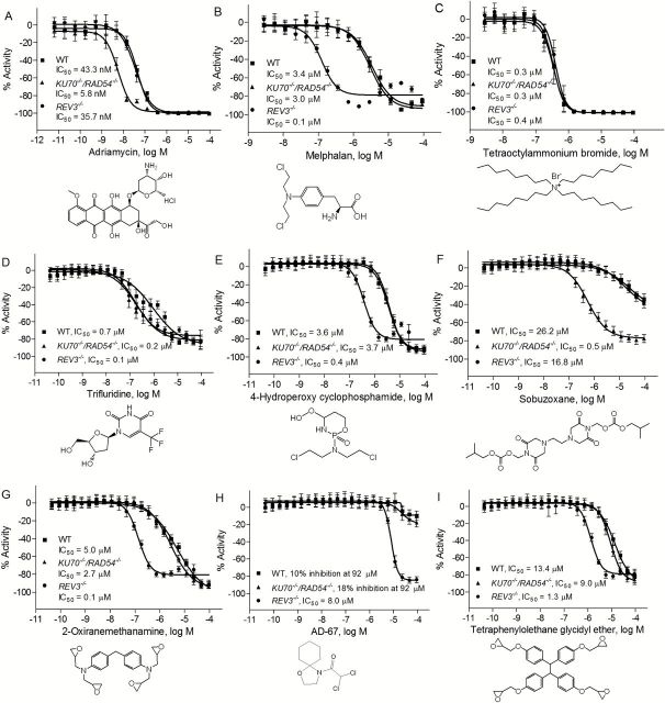
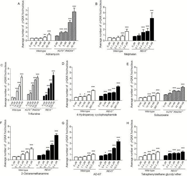
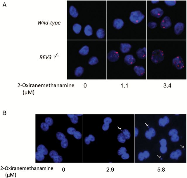
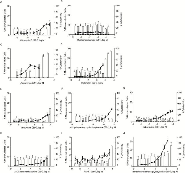
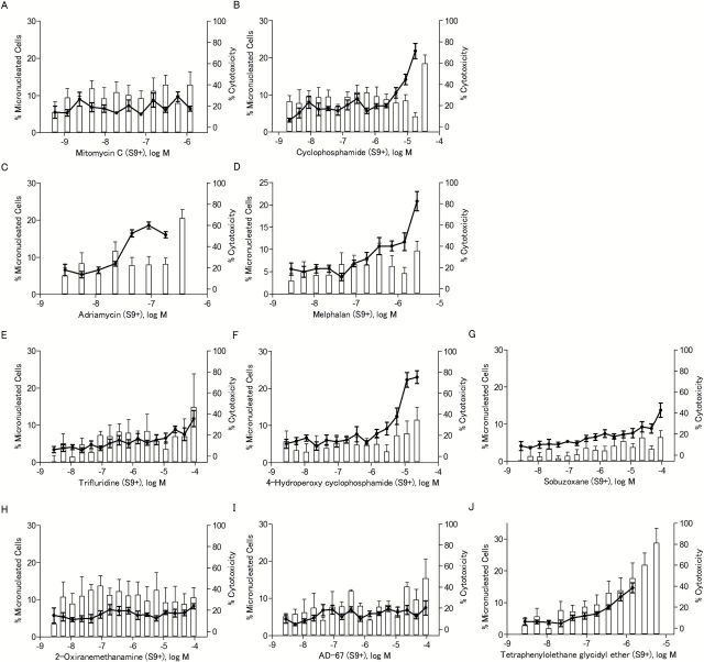
DNA repair pathways play a critical role in maintaining cellular homeostasis by repairing DNA damage induced by endogenous processes and xenobiotics, including environmental chemicals. Induction of DNA damage may lead to genomic instability, disruption of cellular homeostasis and potentially tumours. Isogenic chicken DT40 B-lymphocyte cell lines deficient in DNA repair pathways can be used to identify genotoxic compounds and aid in characterising the nature of the induced DNA damage. As part of the US Tox21 program, we previously optimised several different DT40 isogenic clones on a high-throughput screening platform and confirmed the utility of this approach for detecting genotoxicants by measuring differential cytotoxicity in wild-type and DNA repair-deficient clones following chemical exposure. In the study reported here, we screened the Tox21 10K compound library against two isogenic DNA repair-deficient DT40 cell lines (KU70 (-/-) /RAD54 (-/-) and REV3 (-/-) ) and the wild-type cell line using a cell viability assay that measures intracellular adenosine triphosphate levels. KU70 and RAD54 are genes associated with DNA double-strand break repair processes, and REV3 is associated with translesion DNA synthesis pathways. Active compounds identified in the primary screening included many well-known genotoxicants (e.g. adriamycin, melphalan) and several compounds previously untested for genotoxicity. A subset of compounds was further evaluated by assessing their ability to induce micronuclei and phosphorylated H2AX. Using this comprehensive approach, three compounds with previously undefined genotoxicity-2-oxiranemethanamine, AD-67 and tetraphenylolethane glycidyl ether-were identified as genotoxic. These results demonstrate the utility of this approach for identifying and prioritising compounds that may damage DNA.


Figures

Figure 1.
Figure 1.
Flow chart for the identification of genotoxic compounds. One hundred and nineteen compounds with ≥3-fold increase in cytotoxicity (P < 0.05) in the KU70 −/−/RAD54 −/− and/or REV3 −/− cells compared with wild-type cells were identified in the primary screening. Sixty-three of the 119 compounds were confirmed in a replicate qHTS cells viability assay using the same 3-fold differential cytotoxicity measure. Eight compounds were selected from the 63 based on their novelty, commercial availability and potency of the differential cytotoxicity response for further testing in in vitro γH2AX immunostaining and MN assays.
Figure 2.
Figure 2.
Confirmation of genotype of DT40 clones that were used in this study. (A) Southern blot analysis of indicated gene disrupted DT40 clones using the gene specific probes. (B) Liquid survival assay after exposure to ionising radiation. Cells were cultured for 48h after IR. Cellular ATP level was used to measure cell survival. The survival of untreated cells was set as 100%. (C) Colony survival assay after exposure to IR. Percent survival was determined relative to numbers of colonies from untreated cells. For both B and C, error bars represent SD from at least three independent experiments.
Figure 3.
Figure 3.
Sensitivity of DT40 DNA repair-deficient and the parental cell lines to chemical compounds selected from the primary screening. Cellular survival was determined using CellTiter-Glo after 40h exposure to adriamycin (positive control) (A), melphalan (positive control) (B), tetraoctylammonium bromide (C), trifluridine (D), 4-hydroperoxy cyclophosphamide (E), sobuzoxane (F), 2-oxiranemethanamine (G), AD-67 (H) and tetraphenylolethane glycidyl ether (I). Error bars represent SD from at least three independent experiments.
Figure 4.
Figure 4.
Induction of γH2AX foci in wild-type and DNA repair-deficient DT40 clones after 24h chemical treatment. The y-axis represents the average number of γH2AX foci per nucleus from three independent experiments. Error bars represent SEM. *P < 0.05, **P < 0.01 and ***P < 0.001.
Figure 5.
Figure 5.
Generation of γH2AX foci in DT40 cells and micronucleus formation in CHO-K1 cells treated with 2-oxiranemethanamine. (A) Dose-dependent γH2AX foci (red) formation in DT40 cells. The indicated DT40 clones were treated with 2-oxiranemethanamine for 24h. Images were acquired in ImageXpress using a 40× objective. Hoechst staining (blue) indicates DNA. (B) Micronucleus formation in CHO-K1 cells. CHO-K1 cells were treated with 2-oxiranemethanamine for 24h without S9 treatment. Hoechst staining (blue) indicates DNA. Images were acquired in ImageXpress using a 20× objective. Arrows indicate a cell with MN.
Figure 6.
Figure 6.
The frequency of micronucleated cells after chemical treatment in the absence of S9. The line chart represents the percentage of binucleated cells with MN out of the total number of binucleated cells evaluated. The bar chart represents the NDI-based cytotoxicity. Data represents the mean ± SEM from three experiments.
Figure 7.
Figure 7.
The frequency of micronucleated cells after chemical treatment in the presence of S9. The line chart represents the percentage of binucleated cells with MN out of the total number of binucleated cells evaluated. The bar chart represents the NDI-based cytotoxicity. Data represents the mean ± SEM from three experiments.

References

    1. Ciccia A., Elledge S. J. (2010) The DNA damage response: making it safe to play with knives. Mol. Cell, 40, 179–204. - PMC - PubMed
    1. Hendriks G., van de Water B., Schoonen W., Vrieling H. (2013) Cellular-signaling pathways unveil the carcinogenic potential of chemicals. J. Appl. Toxicol., 33, 399–409. - PubMed
    1. Yamamoto K. N., Hirota K., Kono K., et al. (2011) Characterization of environmental chemicals with potential for DNA damage using isogenic DNA repair-deficient chicken DT40 cell lines. Environ. Mol. Mutagen., 52, 547–561. - PMC - PubMed
    1. Attene-Ramos M. S., Miller N., Huang R., et al. (2013) The Tox21 robotic platform for the assessment of environmental chemicals—from vision to reality. Drug Discov. Today, 18, 716–723. - PMC - PubMed
    1. Tice R. R., Austin C. P., Kavlock R. J., Bucher J. R. (2013) Improving the human hazard characterization of chemicals: a Tox21 update. Environ. Health Perspect., 121, 756–765. - PMC - PubMed

Publication types