. 2015 Jul 24:8:1823-30.
doi: 10.2147/OTT.S86502. eCollection 2015.

Follicular dendritic cell sarcoma: two rare cases and a brief review of the literature

Affiliations

Follicular dendritic cell sarcoma: two rare cases and a brief review of the literature

Yuan Ma et al. Onco Targets Ther. .

Abstract

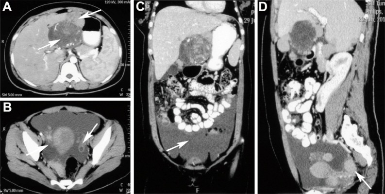
Follicular dendritic cell sarcoma (FDCS) is a rare malignant tumor recognized in recent years. It accounts for only 0.4% of soft-tissue sarcomas, and its underlying causes are largely unknown. A correct diagnosis can be difficult to make. Diagnosis of FDCS depends on the combined clinical examination, histopathologic features, electron microscopic examination and confirmation with immunohistochemical studies. Here, we report two rare cases of FDCS: one case involving multiple bones, and the other involving extensive abdominal and pelvic cavities. Clinical, histopathological, and immunohistochemical aspects, therapeutic options, and a related literature review of the two cases are discussed. As the prevalence of FDCS is increasing, the details of these rare cases may highlight the importance and facilitate treatment of similar diseases.

Keywords: FDCS; abdominal cavity; bone; diagnosis; pelvic cavity; therapy.


Figures

Figure 1
Figure 1
The whole-body bone imaging of case 1. Note: Imaging demonstrated high levels of radioactivity accumulation in multiple bones, such as the sternum, spine, ribs, shoulder blade, and others. The first set of images (A) mainly showed sternum, spine, and pelvis metastases. The second set of images (B) mainly showed shoulder blades, ribs and femur metastases. Abbreviations: L, left; R, right.
Figure 2
Figure 2
Computed tomography images of case 2. Notes: (A) Located in gastric omental bursa, low attenuation mass (arrow) showed heterogeneous enhancement on arterial phase CT. Nodular thickening of the liver capsule could be seen (arrow). (B) CT image of the pelvic cavity showed cystic-solid mixed masses (arrow) in bilateral annex area. (C) Coronal postcontrast CT scan showed a large amount of fluid (arrow) in the abdominal and pelvic cavity. (D) Sagittal postcontrast CT scan showed multiple nodules (arrow) in the pelvic peritoneum area. Abbreviation: CT, computed tomography.
Figure 3
Figure 3
Tumor histology of case 1. Notes: (A) The histopathological appearance indicated that the tumor was composed of cells arranged in a wispy and storiform pattern and that these cells were admixed with lymphocytes (hematoxylin and eosin stain, ×200). (B) Positive for CD21 staining (×200). (C) Negative for CD23 staining (×200). (D) Positive for CD35 staining (×200). (E) Negative for CD68 staining (×200). (F) Negative for CD45 staining (×200). (G) Negative for D2-40 staining (×200). (H) Negative for EBER staining (×200). Abbreviation: EBER, Epstein–Barr virus-encoded RNA.
Figure 4
Figure 4
Tumor histology of case 2. Notes: (A) Section demonstrated oval and long spindle cells arranged in bundles (hematoxylin and eosin stain, ×200). (B) Positive immunohistochemical staining for CD21 (×200). (C) Positive immunohistochemical staining for CD23 (×200). (D) Positive immunohistochemical staining for CD35 (×200). (E) Negative for CD68 staining (×200). (F) Negative for CD45 staining (×200). (G) Negative for D2-40 staining (×200). (H) Negative for EBER staining (×200). Abbreviation: EBER, Epstein–Barr virus-encoded RNA.

References

    1. van Nierop K, de Groot C. Human follicular dendritic cells: function, origin and development. Semin Immunol. 2002;14(4):251–257. - PubMed
    1. Karabulut B, Orhan KS, Guldiken Y, et al. Follicular dendritic cell sarcoma of the nasopharynx. Int J Oral Maxillofac Surg. 2012;41(2):218–220. - PubMed
    1. Monda L, Warnke R, Rosai J. A primary lymph node malignancy with features suggestive of dendritic reticulum cell differentiation. A report of 4 cases. Am J Pathol. 1986;122(3):562. - PMC - PubMed
    1. Chan JK, Fletcher CD, Nayler SJ, et al. Follicular dendritic cell sarcoma. Clinicopathologic analysis of 17 cases suggesting a malignant potential higher than currently recognized. Cancer. 1997;79(2):294–313. - PubMed
    1. Grogg KL, Lae ME, Kurtin PJ, et al. Clusterin expression distinguishes follicular dendritic cell tumors from other dendritic cell neoplasms: report of a novel follicular dendritic cell marker and clinicopathologic data on 12 additional follicular dendritic cell tumors and 6 additional interdigitating dendritic cell tumors. Am J Surg Pathol. 2004;28(8):988–998. - PubMed