

The Use of Biologic Scaffolds in the Treatment of Chronic Nonhealing Wounds
- PMID: 26244105
- PMCID: PMC4505760
- DOI: 10.1089/wound.2014.0604
The Use of Biologic Scaffolds in the Treatment of Chronic Nonhealing Wounds
Abstract
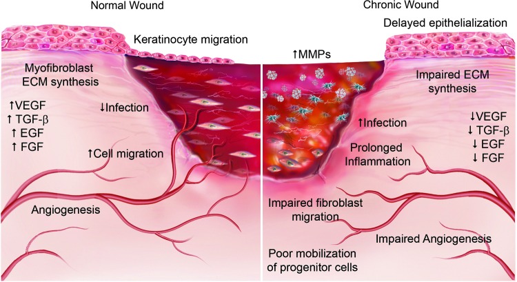
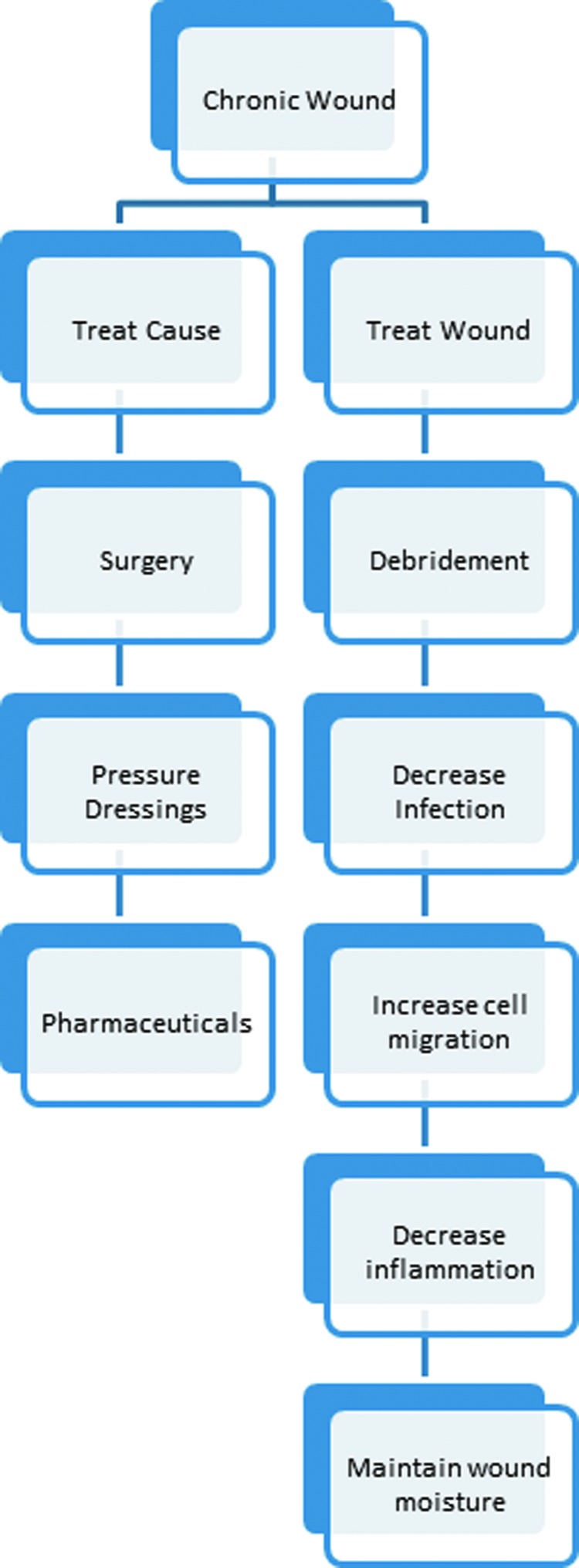
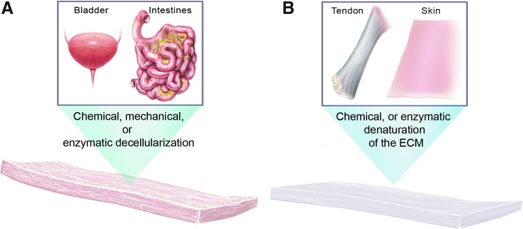
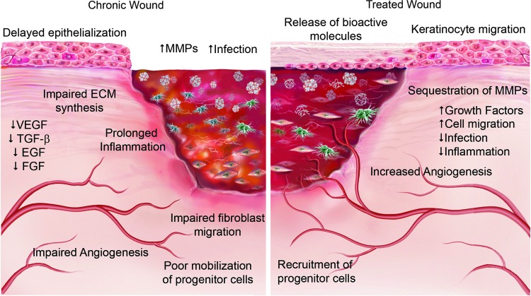
Significance: Injuries to the skin as a result of illness or injury, particularly chronic nonhealing wounds, present a major healthcare problem. Traditional wound care approaches attempt to control the underlying causes, such as infection and ischemia, while the application of wound dressings aims to modify a poorly healing wound environment into a microenvironment more closely resembling an acute wound allowing the body to heal the wound naturally. Recent Advances: Regenerative medicine approaches, such as the use of biologic scaffold materials comprising an intact extracellular matrix (ECM) or individual components of the ECM, are providing new therapeutic options that focus upon the provision of biochemical cues that alter the wound microenvironment to facilitate rapid restoration of normal skin architecture. Critical Issues: The incidence of chronic nonhealing wounds continues to increase. For example, between 15% and 20% of diabetics are likely to develop chronic, nonhealing foot wounds creating an increasing burden on healthcare systems worldwide. Future Directions: Developing a thorough understanding of wound microenvironment and the mechanisms by which biologic scaffolds work in vivo has the potential to markedly improve outcomes in the clinical translation for the treatment of chronic wounds.
Figures

Similar articles
-
Extracellular Matrices (ECM) for Tissue Repair.Surg Technol Int. 2016 Apr;28:43-57. Surg Technol Int. 2016. PMID: 27175813 Review.
-
The Effect of pH on the Extracellular Matrix and Biofilms.Adv Wound Care (New Rochelle). 2015 Jul 1;4(7):431-439. doi: 10.1089/wound.2014.0538. Adv Wound Care (New Rochelle). 2015. PMID: 26155386 Free PMC article. Review.
-
Topical Collagen-Based Biomaterials for Chronic Wounds: Rationale and Clinical Application.Adv Wound Care (New Rochelle). 2016 Jan 1;5(1):19-31. doi: 10.1089/wound.2014.0595. Adv Wound Care (New Rochelle). 2016. PMID: 26858912 Free PMC article. Review.
-
Choosing a Wound Dressing Based on Common Wound Characteristics.Adv Wound Care (New Rochelle). 2016 Jan 1;5(1):32-41. doi: 10.1089/wound.2014.0586. Adv Wound Care (New Rochelle). 2016. PMID: 26858913 Free PMC article. Review.
-
Recent advances in acellular regenerative tissue scaffolds.Clin Podiatr Med Surg. 2015 Jan;32(1):147-59. doi: 10.1016/j.cpm.2014.09.008. Clin Podiatr Med Surg. 2015. PMID: 25440425 Review.
Cited by
-
Exosomes from adipose-derived stem cells and application to skin wound healing.Cell Prolif. 2021 Mar;54(3):e12993. doi: 10.1111/cpr.12993. Epub 2021 Jan 17. Cell Prolif. 2021. PMID: 33458899 Free PMC article. Review.
-
Functional Properties of a Purified Reconstituted Bilayer Matrix Design Support Natural Wound Healing Activities.Plast Reconstr Surg Glob Open. 2021 May 21;9(5):e3596. doi: 10.1097/GOX.0000000000003596. eCollection 2021 May. Plast Reconstr Surg Glob Open. 2021. PMID: 34036030 Free PMC article.
-
Engineered Biopolymeric Scaffolds for Chronic Wound Healing.Front Physiol. 2016 Aug 5;7:341. doi: 10.3389/fphys.2016.00341. eCollection 2016. Front Physiol. 2016. PMID: 27547189 Free PMC article. Review.
-
Effectiveness of fire needle combining with moist healing dressing to promote the growth of granulation tissue in chronic wounds: A case report.Int J Nurs Sci. 2020 May 30;7(3):386-390. doi: 10.1016/j.ijnss.2020.05.008. eCollection 2020 Jul 10. Int J Nurs Sci. 2020. PMID: 32817864 Free PMC article.
-
Novel application of absorbable gelatine sponge combined with polyurethane film for dermal reconstruction of wounds with bone or tendon exposure.Int Wound J. 2023 Jan;20(1):18-27. doi: 10.1111/iwj.13832. Epub 2022 May 5. Int Wound J. 2023. PMID: 35510525 Free PMC article.
References
-
- Cherry DK, Hing E, Woodwell DA, Rechtsteiner EA. National Ambulatory Medical Care Survey: 2006 summary. Natl Health Stat Report 1–31, 2008 - PubMed
-
- Lawrence WT, Diegelmann RF. Growth factors in wound healing. Clin Dermatol 1994;12:157–169 - PubMed
-
- Centers for Disease Control and Prevention. National diabetes fact sheet: general information and national estimates on diabetes in the United States, 2007. Atlanta, GA: Department of Health and Human Services, 2008
-
- Reiber GE, Boyko EJ, Smith DG. Lower-extremity foot ulcers and amputations in diabetes. In: Group NDD, ed. Diabetes in America, Second Edition. Washington, DC: National Institutes of Health, 1995:409–428
-
- Gurtner GC, Werner S, Barrandon Y, Longaker MT. Wound repair and regeneration. Nature 2008;453:314–321 - PubMed
Publication types
LinkOut - more resources
Full Text Sources
Other Literature Sources
Medical
