Review
. 2015 Jul 16;3(7):556-74.
doi: 10.12998/wjcc.v3.i7.556.

From variome to phenome: Pathogenesis, diagnosis and management of ectopic mineralization disorders

Affiliations
Review

From variome to phenome: Pathogenesis, diagnosis and management of ectopic mineralization disorders

Eva Yg De Vilder et al. World J Clin Cases. .

Abstract

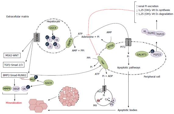
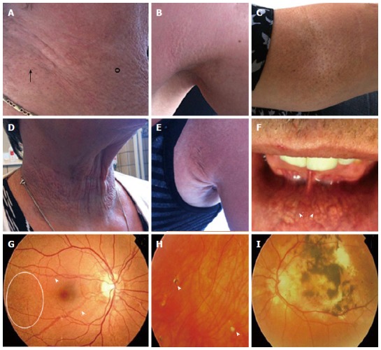
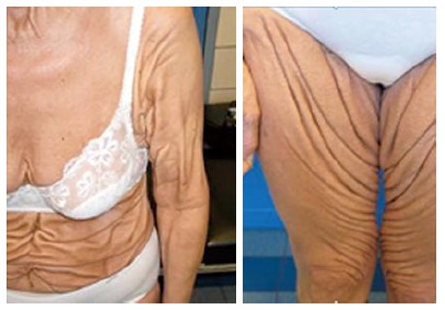
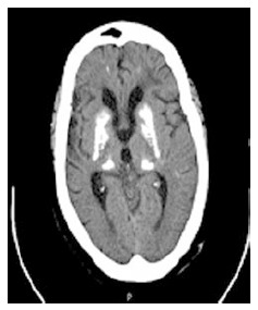
Ectopic mineralization - inappropriate biomineralization in soft tissues - is a frequent finding in physiological aging processes and several common disorders, which can be associated with significant morbidity and mortality. Further, pathologic mineralization is seen in several rare genetic disorders, which often present life-threatening phenotypes. These disorders are classified based on the mechanisms through which the mineralization occurs: metastatic or dystrophic calcification or ectopic ossification. Underlying mechanisms have been extensively studied, which resulted in several hypotheses regarding the etiology of mineralization in the extracellular matrix of soft tissue. These hypotheses include intracellular and extracellular mechanisms, such as the formation of matrix vesicles, aberrant osteogenic and chondrogenic signaling, apoptosis and oxidative stress. Though coherence between the different findings is not always clear, current insights have led to improvement of the diagnosis and management of ectopic mineralization patients, thus translating pathogenetic knowledge (variome) to the phenotype (phenome). In this review, we will focus on the clinical presentation, pathogenesis and management of primary genetic soft tissue mineralization disorders. As examples of dystrophic calcification disorders Pseudoxanthoma elasticum, Generalized arterial calcification of infancy, Keutel syndrome, Idiopathic basal ganglia calcification and Arterial calcification due to CD73 (NT5E) deficiency will be discussed. Hyperphosphatemic familial tumoral calcinosis will be reviewed as an example of mineralization disorders caused by metastatic calcification.

Keywords: Arterial calcification due to CD73 deficiency; Ectopic mineralization; Etiology; Generalized arterial calcification of infancy; Hyperphosphatemic familial tumoral calcinosis; Idiopathic basal ganglia calcification; Keutel syndrome; Phenotype; Pseudoxanthoma elasticum; Pseudoxanthoma elasticum-like syndrome.


Figures

Figure 1
Figure 1
Schematic representation of the pathophysiological mechanisms leading to ectopic mineralization. Hepatocyte: Impairment of ABCC6 function leads to upregulation of pro-osteogenic pathways (MSX2-WNT, TGFβ-Smad 2/3, BMP2-Smad-RUNX2), upregulation of their downstream targets and eventually to ectopic mineralization. GGCX carboxylates and hence activates multiple targets, such as coagulation factors and MGP, the latter being a potent BMP2-inhibitor and hence mineralization inhibitor. When GGCX function is impaired, these targets stay inactive, leading to increased mineralization. ENPP1 converts ATP to AMP and PPi, the latter being a mineralization inhibitor. Impairment of this conversion and hence a decrease in the PPi level leads to increase in ectopic mineralization. Peripheral cell: After glycosylation by GALNT3, FGF23 forms a complex with FGFR1 and KL (coreceptor) which leads to increased renal excretion of Pi, a pro-mineralizing agent and decreased 1,25 dihydroxyvitamin D3, causing a decrease in intestinal Pi absorption. NT5E converts AMP to Pi and adenosine, which inhibits the pro-mineralizing TNAP. Impairment of NT5E function leads to increased TNAP activity and decreased PPi concentration, hence leading to ectopic mineralization. Pi is internalized into the peripheral cell by PiT2 and leaves the cell through apoptotic bodies, which cause ectopic mineralization through apoptotic pathways (not shown). In MVs an influx occurs of Pi via PiT2 and of Ca2+, which is facilitated by A and PS. This leads to an accumulation of growing hydroxyapatite crystals, eventually causing the MVs to burst and the crystals to grow in the extracellular matrix. A: Annexin A5; ABCC6: Adenosine triphosphate-binding cassette, subfamily C, member 6; ADP: Adenosine diphosphate; AMP: Adenosine monophosphate; ATP: Adenosine triphosphate; BMP2: Bone morphogenetic protein 2; C: Carboxyl; Ca2+: Calcium 2+; ENPP1: Ectonucleotide pyrophosphatase/phosphodiesterase 1; FGF23: Fibroblast growth factor 23; FGFR1: Fibroblast growth factor receptor 1; G: Glycosyl-; GALNT3: UDP-N-acetyl-alpha-D-galactosamine: Polypeptide N-acetylgalactosaminyltransferase 3; GGCX: Gamma-glutamyl carboxylase; HA: Hydroxyapatite; KL: Klotho; MGP: Matrix gla protein; MMP9: Matrix metalloproteinase; MSX2: Muscle segment homeobox, drosophila, homolog of, 2; MV: Matrix vesicle; NT5E: Ecto-5-prime nucleotidase or CD73; OC: Osteocalcine; Pi: Inorganic phosphate; SLC20A2: Solute carrier family 20 (phosphate transporter), member 2; PPi: Inorganic pyrophosphate; PS: Phosphatidyl serine; RUNX2: Runt-related transcription factor; Smad: Mothers against decapentaplegic, drosophila, homolog of; TGFβ: Transforming growth factor β; TNAP: Tissue-non-specific alkaline phosphatase; VEGF: Vascular endothelial growth factor; WNT: Wingless-type MMTV integration site family; II, VII, IX, X: Vitamin K-dependent coagulation factors; 1,25 (OH)2 Vit D3: 1,25-dihydroxyvitamine D3 (calcitriol).
Figure 2
Figure 2
Dermatological (A-F) and ophthalmological (G-I) manifestations of pseudoxanthoma elasticum. A, B: Flexural areas can show papular lesions (°) and coalesced plaques of papules (arrow); C: Cutaneous peau d’orange; D, E: Additional skin folds; F: Yellowish, reticular pattern on the mucosae of the lip (arrowed); G: Ocular fundi show peau d’orange (circle) and angioid streaks (arrowed); H: Comets and comet tails (arrowhead); I: Choroidal and subretinal hemorrhage.
Figure 3
Figure 3
Cutaneous features of a pseudoxanthoma elasticum-like patient with increased amount of generalized thick leathery skin folds.
Figure 4
Figure 4
Transverse computed tomography of the brain displaying symmetrical bilateral ganglia calcification in an idiopathic basal ganglia calcification patient.

Similar articles

Cited by

References

    1. Vanakker O, Hosen MJ, De Paepe A. Ectopic soft tissue calcification: process, determinants and health impact. Hauppauge, NY: Human Anatomy and Physiology; 2013. pp. 261–304.
    1. Kirsch T. Biomineralization--an active or passive process? Connect Tissue Res. 2012;53:438–445. - PubMed
    1. Golub EE. Biomineralization and matrix vesicles in biology and pathology. Semin Immunopathol. 2011;33:409–417. - PMC - PubMed
    1. Giachelli CM. Ectopic calcification: gathering hard facts about soft tissue mineralization. Am J Pathol. 1999;154:671–675. - PMC - PubMed
    1. Kornak U. Animal models with pathological mineralization phenotypes. Joint Bone Spine. 2011;78:561–567. - PubMed