. 2015 Aug 5:5:12754.
doi: 10.1038/srep12754.

Crystal structure of E. coli endonuclease V, an essential enzyme for deamination repair

Affiliations

Crystal structure of E. coli endonuclease V, an essential enzyme for deamination repair

Zhemin Zhang et al. Sci Rep. .

Abstract

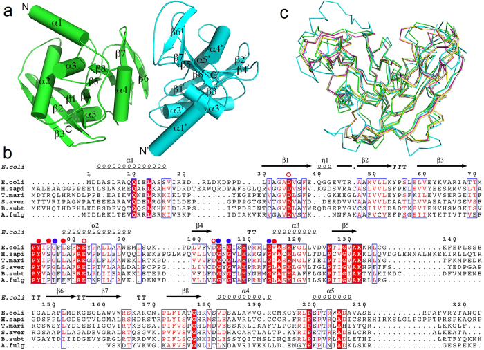
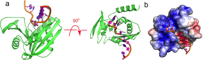
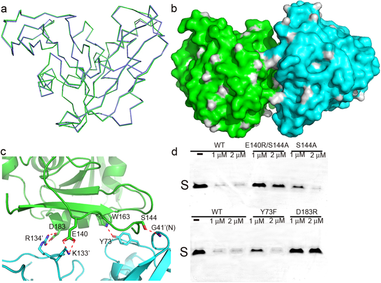
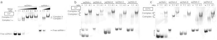
Endonuclease V (EndoV) is a ubiquitous protein present in all three kingdoms of life, responsible for the specific cleavages at the second phosphodiester bond 3' to inosine. E. coli EndoV (EcEndoV) is the first member discovered in the EndoV family. It is a small protein with a compact gene organization, yet with a wide spectrum of substrate specificities. However, the structural basis of its substrate recognition is not well understood. In this study, we determined the 2.4 Å crystal structure of EcEndoV. The enzyme preserves the general 'RNase H-like motif' structure. Two subunits are almost fully resolved in the asymmetric unit, but they are not related by any 2-fold axes. Rather, they establish "head-to-shoulder" contacts with loose interactions between each other. Mutational studies show that mutations that disrupt the association mode of the two subunits also decrease the cleavage efficiencies of the enzyme. Further biochemical studies suggest that EcEndoV is able to bind to single-stranded, undamaged DNA substrates without sequence specificity, and forms two types of complexes in a metal-independent manner, which may explain the wide spectrum of substrate specificities of EcEndoV.


Figures

Figure 1
Figure 1. The structural characterization of EcEndoV.
(a) The overall structure of EcEndoV-SF. The two subunits are colored in cyan and in green. The major secondary elements and the N-, C-termini are labeled. (b) Multiple sequence alignment of E. coli, H. sapiens, T. maritima, S. avermitilis, B. subtilis, A. fulgidus EndoV sequences. The full-length EcEndoV sequence is shown and the secondary structure is drawn on the top by ESPRIPT according to the final structure of EcEndoV-SF. The red solid dots represent the hydrophobic residues that form the hypoxanthine-binding pocket, while the blue dots represent the invariant glycine residues that form backbone hydrogen bonds with the base. The open dots are the Mg2+-binding residues. Residues identical in all four sequences are highlighted in red while similar residues in blue boxes. (c) Structure overlay of Cα traces of EcEndoV-SF (green), hEndoV (cyan, PDB code 4NSP), TmEndoV in the DNA-bound form (magenta, PDB code 2W35), TmEndoV in the DNA-bound form (yellow, PDB code 2W36), TmEndoV in the DNA-bound form (gray, PDB code 4B20). All the DNA components were omitted in the DNA-containing complexes.
Figure 2
Figure 2. The model of EcEndoV-SF-DNA complex in the stoichiometry of 1:1.
(a) Two orthogonal views of the complex in the ribbon form, in which the protein and DNA are shown in green and orange, respectively. DNA coordinates were taken from PDB 2W35 and the model was generated by superposition of the protein model of 2W35 onto apo EcEndoV-SF. (b) The complex with apo EcEndoV-SF in the surface charge distribution. Surface charge potential is calculated by APBS.
Figure 3
Figure 3. The organization pattern of the two subunits in the asymmetric unit.
The color scheme is the same as in Fig. 1a. (a) Superposition of chain A backbone onto that of chain B. (b) The two subunits shown in surface representation. (c) The close-up view of the interactions between the two monomers with the hydrogen bonds indicated by the red dotted lines. (d) The cleavage assay of the mutants that disrupt the association mode. The gels were cropped to reduce the size of the figure. Three single mutants S144A, Y73F, D183R and the double mutant E140R/S144A were tested along with WT EcEndoV, with indicated concentrations. The substrate bands are labeled as “S” and the products are too short to be stained by ethidium bromide.
Figure 4
Figure 4. The formation of two types of complexes and the binding behavior of EcEndoV.
(a) The EMSA of EcEndoV-ssDNA-I/ssRNA-I complexes formed by a constant amount of ssDNA-I, or ssRNA-I (4 μM each) and increasing amounts of protein (molar ratios from 1:1 to 16:1 for ssDNA-I, and 1:1 to 4:1 for ssRNA-I) in the presence of 5 mM Ca2+. The two types of complexes and free probes are indicated on each side. (b) EMSA of the binding of EcEndoV to nonspecific ssDNA in the presence of 5 mM Ca2+. (c) EMSA of the binding of EcEndoV to nonspecific ssDNA in the presence of 2.5 mM EDTA.

References

    1. Shapiro R. & Pohl S. H. The reaction of ribonucleosides with nitrous acid. Side products and kinetics. Biochemistry 7, 448–455 (1968). - PubMed
    1. Shapiro R. & Shiuey S. J. Reaction of nitrous acid with alkylaminopurines. Biochimica et biophysica acta 174, 403–405 (1969). - PubMed
    1. Burney S., Caulfield J. L., Niles J. C., Wishnok J. S. & Tannenbaum S. R. The chemistry of DNA damage from nitric oxide and peroxynitrite. Mutation Research/Fundamental and Molecular Mechanisms of Mutagenesis 424, 37–49, doi: http://dx.doi.org/10.1016/S0027-5107(99)00006-8 (1999). - DOI - PubMed
    1. Dedon P. C. & Tannenbaum S. R. Reactive nitrogen species in the chemical biology of inflammation. Archives of biochemistry and biophysics 423, 12–22 (2004). - PubMed
    1. Lindahl T. Instability and decay of the primary structure of DNA. Nature 362, 709–715, 10.1038/362709a0 (1993). - DOI - PubMed

Publication types

MeSH terms

Substances

LinkOut - more resources