. 2015 Aug 5;10(8):e0134922.
doi: 10.1371/journal.pone.0134922. eCollection 2015.

Enhanced B-Cell Receptor Recognition of the Autoantigen Transglutaminase 2 by Efficient Catalytic Self-Multimerization

Affiliations

Enhanced B-Cell Receptor Recognition of the Autoantigen Transglutaminase 2 by Efficient Catalytic Self-Multimerization

Jorunn Stamnaes et al. PLoS One. .

Abstract

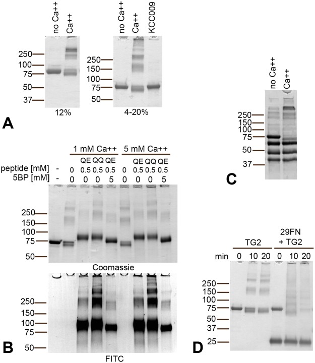
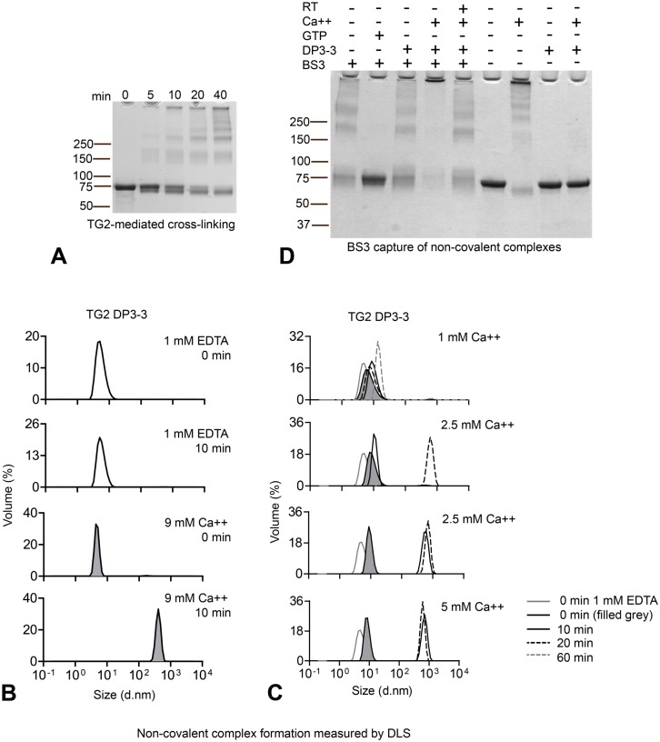
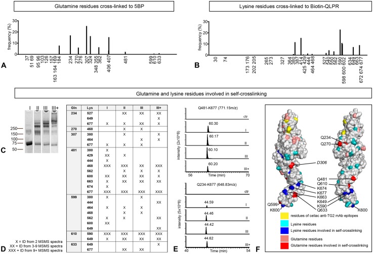
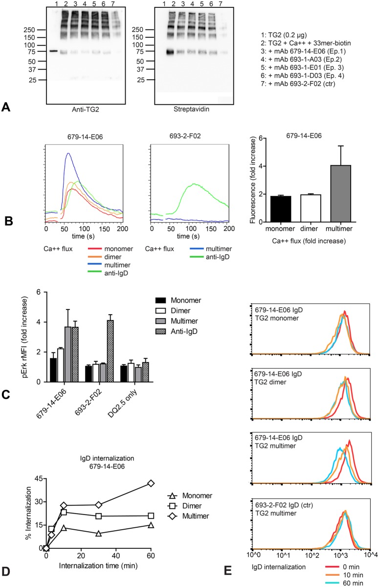
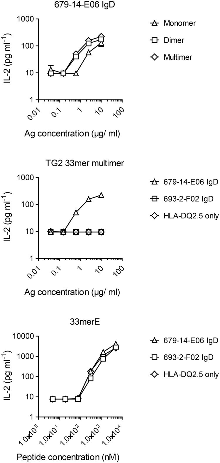
A hallmark of the gluten-driven enteropathy celiac disease is autoantibody production towards the enzyme transglutaminase 2 (TG2) that catalyzes the formation of covalent protein-protein cross-links. Activation of TG2-specific B cells likely involves gluten-specific CD4 T cells as production of the antibodies is dependent on disease-associated HLA-DQ allotypes and dietary intake of gluten. IgA plasma cells producing TG2 antibodies with few mutations are abundant in the celiac gut lesion. These plasma cells and serum antibodies to TG2 drop rapidly after initiation of a gluten-free diet, suggestive of extrafollicular responses or germinal center reactions of short duration. High antigen avidity is known to promote such responses, and is also important for breakage of self-tolerance. We here inquired whether TG2 avidity could be a feature relevant to celiac disease. Using recombinant enzyme we show by dynamic light scattering and gel electrophoresis that TG2 efficiently utilizes itself as a substrate due to conformation-dependent homotypic association, which involves the C-terminal domains of the enzyme. This leads to the formation of covalently linked TG2 multimers. The presence of exogenous substrate such as gluten peptide does not inhibit TG2 self-cross-linking, but rather results in formation of TG2-TG2-gluten complexes. The celiac disease autoantibody epitopes, clustered in the N-terminal part of TG2, are conserved in the TG2-multimers as determined by mass spectrometry and immunoprecipitation analysis. TG2 multimers are superior to TG2 monomer in activating A20 B cells transduced with TG2-specific B-cell receptor, and uptake of TG2-TG2-gluten multimers leads to efficient activation of gluten-specific T cells. Efficient catalytic self-multimerization of TG2 and generation of multivalent TG2 antigen decorated with gluten peptides suggest a mechanism by which self-reactive B cells are activated to give abundant numbers of plasma cells in celiac disease. Importantly, high avidity of the antigen could explain why TG2-specific plasma cells show signs of an extrafollicular generation pathway.


Conflict of interest statement

Competing Interests: The authors have declared that no competing interests exist.

Figures

Fig 1
Fig 1. TG2 self-crosslinking in the absence or presence of competitor substrate.
(A) TG2 (2 μM) incubated in the presence of CaCl2 will cross-link itself into complexes that can be resolved by gradient SDS-PAGE (4–20%) but not by standard 12% SDS-PAGE. Self-crosslinking requires TG2 catalytic activity and does not happen in the absence of CaCl2 or presence of an active site inhibitor. (B) TG2 (1 μM) self-crosslinking as seen by gradient SDS-PAGE (at 15min) occurs even in the presence of a high excess of glutamine donor substrate (DQ2.5-glia-α2-QQ-FITC QQ; DQ2.5-glia-α2-QE-FITC, QE) or primary amine (5BP) (Coomassie). Incorporation of the FITC-labeled peptides is observed both in TG2 monomer and TG2 multimers (FITC). (C) Incubation of TG2 (78 kDa) together with human albumin (64 kDa), human IgG (50 kDa + 25 kDa) and ovalbumin (45 kDa) in the presence of CaCl2 results in selective disappearance of the monomeric TG2 band, indicating that self-crosslinking is a specific event. (D) Incubation of TG2 with excess of the known protein substrate 29kDa FN fragment (29FN) results in disappearance of TG2 monomer and incorporation of both TG2 and 29FN into complexes.
Fig 2
Fig 2. Homotypic TG2 association is conformation dependent
(A) TG2 (2.4 μM) self-crosslinking over time occurs via initial formation of dimer and trimer as observed after 5 and 10 min. (B) Inactive, inhibitor bound TG2 produced in E. coli (TG2-DP3-3) was incubated at 37°C in the presence of 1 mM EDTA or 9 mM CaCl2 and aggregation was measured by DLS. The graphs display volume percentage (Y axis) and size as diameter (d. nm) on the X-axis. Incubation with 9 mM CaCl2 at 37°C results in aggregation formation already after 10 min. (C) Titration of CaCl2 was performed at 37°C and non-covalent complex formation was compared at different time-points: 0 min (black line, grey fill), 10 min (black solid line) and 20 min (black dotted line). For 1 mM CaCl2, 60 min incubation is also shown (grey dotted line). The solid grey line shows TG2-DP3-3 incubated in 1 mM EDTA at time 0 min for comparison. Two datasets from the same experiment are shown for 2.5 mM CaCl2 as variation was observed in the degree of high molecular weight multimer formation for this CaCl2 concentration. (D) Capture of non-covalent TG2-TG2 associations using BS3 cross-linking reagent. TG2 was incubated in the absence of effectors or together with 5 mM CaCl2 or 1 mM GTP for 30 min at 37°C or, where indicated, for 30 min at room temperature (RT) before addition of BS3. To avoid enzymatic self-crosslinking in the presence of Ca2+ the enzyme was pretreated with the active-site inhibitor DP3-3. Incubation of TG2-DP3-3 with CaCl2 at 37°C followed by BS3 treatment results in the formation of large complexes that do not migrate through the gel.
Fig 3
Fig 3. The C-terminal domains are required for homotypic association and self-crosslinking
(A) A truncated variant of TG2 lacking the two C-terminal domains (1–465) was expressed in E. coli and treated with DP3-3. The inactive, inhibitor-bound deletion mutant (1-465-DP3-3) was incubated at 37°C in the presence of 1 mM EDTA or 9 mM CaCl2 and aggregation was measured by DLS. The graphs display volume percentage (Y axis) and diameter (d.nm) on the X-axis. No multimer formation of the truncated TG2 was observed following 40 min incubation. (B) Capture of non-covalent complexes of inactive E. coli-produced DP3-3-treated full length (WT) and 1–465 deletion mutant (1–465) TG2 following incubation at 37°C using BS3 as described in Fig 2C. No complex formation was observed for the deletion mutant compared to full-length TG2. (C) Incubation of active E. coli-produced deletion mutant (1–465) with CaCl2 did not result in self-crosslinking as observed for full length E. coli-produced TG2. The gel shows incubation of different enzyme concentrations with 5 mM CaCl2 at 37°C for 60 min: 1) 0.7 μM, 2) 1.4 μM, 4) 4.2 μM and 3) 1.4 μM in the presence of 0.2 mM DQ2.5-glia-α2-QQ-FITC. Incorporated gluten peptides are visualized by FITC fluorescence.
Fig 4
Fig 4. TG2-mediated self-crosslinking sites locate to the catalytic core and C-terminal part of TG2
(A) Glutamine residues of TG2 cross-linked to 5BP. The frequency (%) reflects the abundance of modified residues compared to each other. (B) Lysine residues of TG2 cross-linked to the small peptide Biotin-QLPR. Histograms in (A) and (B) are representative of two and three independent experiments, respectively. (D) Cross-linked peptide adducts identified from in solution tryptic digests of size separated fractions shown in (C). Multiple cross-linked peptide adducts were identified and only peptide adducts identified by two or more independent MSMS scans are listed in (D). Results from the second sample of triplicates are shown. (E) Extracted ion chromatogram for the peptide adducts Q481-K677 (AVKGFR-VGQSMNMGSDFDVFAHITNNTAEEYVC(acetamide)R, 771.15 m/z; EIC window 771.15–771.25 m/z,) and Q234-K677 (AVKGFR-VVSGMVNC(acetamide)NDDQGVLLGR; 648.83m/z, EIC window 648.83–648.93m/z). Signal intensity (y-axis) reflects peptide abundance. (F) Location of glutamine and lysine residues in open conformation TG2 (PDB ID code 2Q3Z). Glutamine residues identified in cross-linked peptide adducts are shown in red, the remaining glutamine residues are shown in pink. Lysine residues identified in cross-linked peptide adducts are shown in blue, the remaining lysine residues are shown in turquoise. Residues E8, R19, K30 and D94, important for recognition of celiac anti-TG2 monoclonal antibodies [26], are shown in yellow. Q307 is not resolved in the crystal structure, and D306 (italics) is highlighted instead. K464 and K468 are also located in unresolved parts.
Fig 5
Fig 5. TG2 multimers are superior to TG2 monomer in activation of TG2-specific A20 B cells
(A) Celiac TG2-specific monoconal antibodies are able to bind TG2-gluten multimer complexes. TG2 crosslinked in the presence of biotin-33mer peptide was immunoprecipitated using TG2-specific monoclonal antibodies representing all four celiac epitopes, or using a non TG2-specific monoclonal antibody (693-2-F02). TG2 was detected with mouse monoclonal antibody CUB7402 and TG2-bound peptide was detected with HRP-conjugated streptavidin. (B) Intracellular Ca2+ flux was compared following stimulation of transduced A20 B cells expressing HLA-DQ2.5 and TG2-specific (679-14-E06) or non TG2-specific (693-2-F02) IgD BCR with size-separated TG2 complexes or bivalent mouse anti-human IgD antibody. TG2 multimers were superior to monomer and dimers. Calcium flux induced by TG2 was BCR dependent and not observed in control cells. (C) A20 cells expressing TG2-specific (679-14-E06), non TG2-specific (693-2-F02) BCR or only HLA-DQ2.5 were incubated with complexes of TG2 and FITC-labeled 33mer peptide or with anti-human IgD antibody followed by staining for intracellular phoshorylated Erk (pErk). ERK phosphorylation was BCR-dependent and was not observed in A20 cells expressing only HLA-DQ2.5. (D, E) A20 cells expressing HLA-DQ2.5 and TG2-specific (679-14-E06) or non TG2-specific (693-2-F02) IgD BCR were incubated on ice with complexes of TG2 followed by incubation at 37°C. BCR internalization was assessed by flow cytometry staining for surface IgD. Columns in (B) show the means of triplicates from the second experiment (+SD), columns in (D) show the means of two independent experiments (+SD).
Fig 6
Fig 6. Presentation of gluten peptide by TG2-specific B cells upon incubation with TG2-gluten peptide complexes
A20 B cells expressing HLA-DQ2.5 alone or in combination with TG2-specific (679-14-E06) or non TG2-specific (693-2-F02) IgD BCR were incubated with size-fractionated complexes of TG2 and FITC-33mer peptide, and their ability to present deamidated peptide to DQ2.5-glia-α2-specific hybridoma T cells was assessed by measuring release of IL-2. When incubated with the free, deamidated peptide (33merE), all A20 cells were capable of presenting peptide to the T cells. Only TG2-specific cells, however, could take up and present complexes of TG2 and peptide.

References

    1. Jabri B, Sollid LM. Tissue-mediated control of immunopathology in coeliac disease. Nat Rev Immunol. 2009;9(12):858–70. 10.1038/nri2670 - DOI - PubMed
    1. Lorand L, Graham RM. Transglutaminases: crosslinking enzymes with pleiotropic functions. Nat Rev Mol Cell Biol. 2003;4(2):140–56. - PubMed
    1. Vader LW, de Ru A, van der Wal Y, Kooy YM, Benckhuijsen W, Mearin ML, et al. Specificity of tissue transglutaminase explains cereal toxicity in celiac disease. J Exp Med. 2002;195(5):643–9. - PMC - PubMed
    1. Fleckenstein B, Molberg O, Qiao SW, Schmid DG, von der Mulbe F, Elgstoen K, et al. Gliadin T cell epitope selection by tissue transglutaminase in celiac disease. Role of enzyme specificity and pH influence on the transamidation versus deamidation process. J Biol Chem. 2002;277(37):34109–16. - PubMed
    1. Dorum S, Arntzen MO, Qiao SW, Holm A, Koehler CJ, Thiede B, et al. The preferred substrates for transglutaminase 2 in a complex wheat gluten digest are peptide fragments harboring celiac disease T-cell epitopes. PLoS ONE. 2010;5(11):e14056 10.1371/journal.pone.0014056 - DOI - PMC - PubMed

Publication types

MeSH terms