. 2015 Jul 15;82(1):e1-e10.
doi: 10.4102/ojvr.v82i1.953.

Thoracic limb morphology of the red panda (Ailurus fulgens) evidenced by osteology and radiography

Affiliations

Thoracic limb morphology of the red panda (Ailurus fulgens) evidenced by osteology and radiography

Modesta Makungu et al. Onderstepoort J Vet Res. .

Abstract

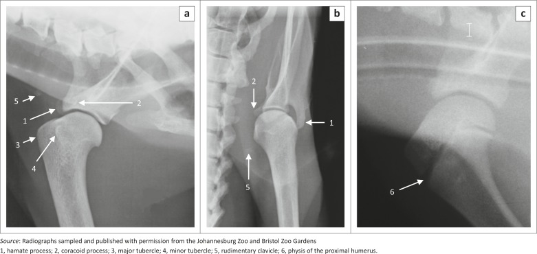
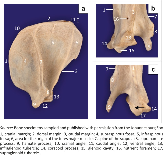
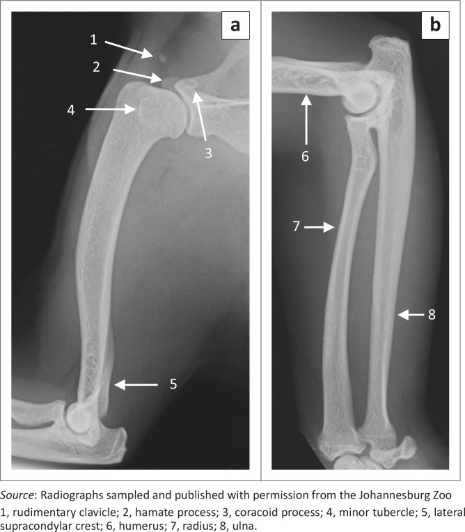
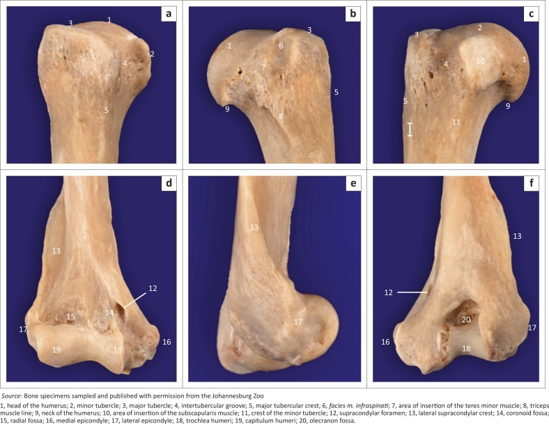
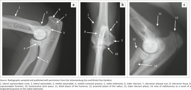
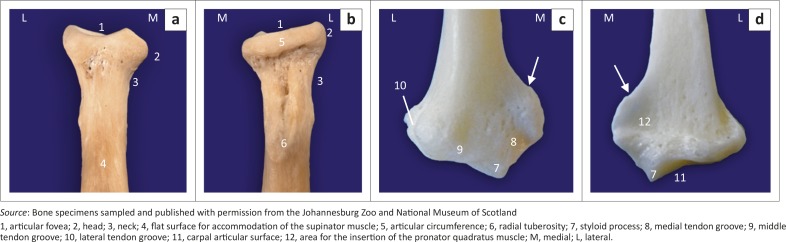
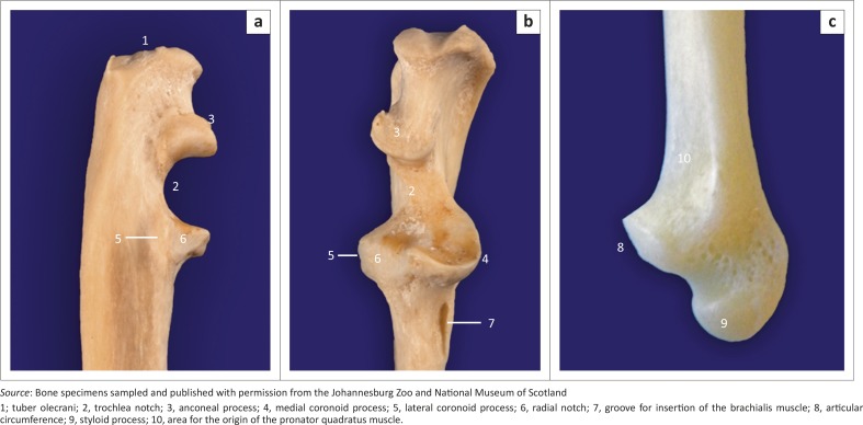
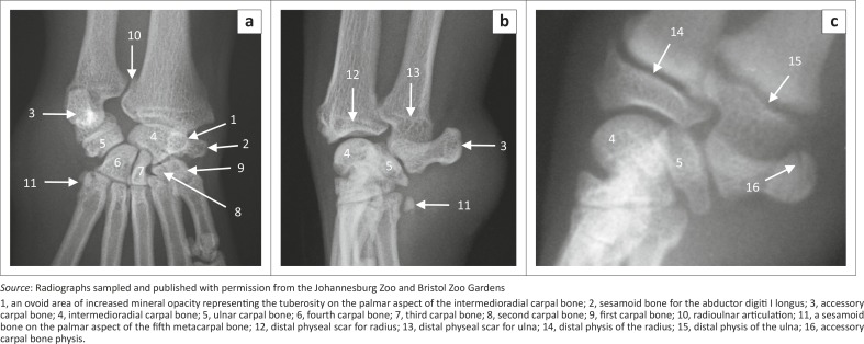
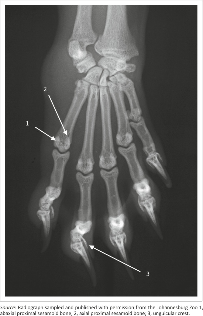
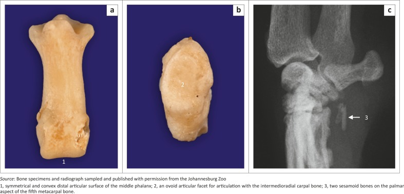
The red panda (Ailurus fulgens) is distributed primarily in the Himalayas and southern China. It is classified as a vulnerable species by the International Union for Conservation of Nature. The aim of this study was to describe the normal osteology and radiographic anatomy of the thoracic limb of the red panda. Radiography of the right thoracic limb was performed in seven captive adult red pandas. Radiographic findings were correlated with bone specimens from three adult animals. The scapula was wide craniocaudally and presented with a large area for the origin of the teres major muscle. The square-shaped major tubercle did not extend proximal to the head of the humerus. The medial epicondyle was prominent. A supracondylar foramen was present. The radial tuberosity and sesamoid bone for the abductor digiti I longus were prominent. The accessory carpal bone was directed palmarolaterally. Metacarpal bones were widely spread. The thoracic limb morphology of the red panda evidenced by osteology and radiography indicated flexibility of the thoracic limb joints and well-developed flexor and supinator muscles, which are important in arboreal quadrupedal locomotion. Knowledge gained during this study may prove useful in identifying skeletal material or remains and diagnosing musculoskeletal diseases and injuries of the thoracic limb.


Conflict of interest statement

The authors declare that they have no financial or personal relationships that may have inappropriately influenced them in writing this article.

Figures

FIGURE 1
FIGURE 1
Radiographs of the right shoulder joints of (a, b) a 14.3-year-old male and (c) a skeletally immature red panda. Mediolateral views are shown in (a) and (c) and a caudocranial view in (b).
FIGURE 2
FIGURE 2
(a) Lateral, (b) caudal and (c) cranial views of a specimen of the right scapula of an adult red panda. Black arrow shows the proximal extension of the articular surface of the glenoid cavity.
FIGURE 3
FIGURE 3
Mediolateral radiographs of (a) the right humerus and (b) the radius and ulna of two male red pandas. Bones of a 1.4-year-old animal are shown in (a) and those of a 2.5-year-old animal are shown in (b).
FIGURE 4
FIGURE 4
Bone specimen of (a–c) the right proximal and (d–f) the distal humerus of an adult red panda. Cranial views are shown in (a) and (d). Lateral views are shown in (b) and (e). Medial and caudal views are shown in (c) and (f), respectively.
FIGURE 5
FIGURE 5
Radiographs of the right elbow joint of (a) a 1.4-year-old, (b) a 10.3-year-old and (c) a skeletally immature male red panda. Mediolateral views are shown in (a) and (c) and a craniocaudal view is shown in (b).
FIGURE 6
FIGURE 6
Bone specimens of (a, b) the right proximal and (c, d) the distal radius of adult red pandas. Cranial views are shown in (a) and (c). Caudal views are shown in (b) and (d). White arrows in (c) and (d) indicate a prominent crest for the insertion of the brachioradialis muscle.
FIGURE 7
FIGURE 7
(a) Lateral, (b) cranial and (c) medial views of bone specimens of (a, b) the right proximal and (c) the distal ulna of adult red pandas.
FIGURE 8
FIGURE 8
(a) Dorsopalmar and (b, c) mediolateral radiographs of the right carpus of red pandas. The carpus of a 1.4-year-old female animal is shown in (a) and (b), whereas that of a skeletally immature animal is shown in (c).
FIGURE 9
FIGURE 9
Dorsopalmar radiograph of the right manus of a 1.4-year-old female red panda.
FIGURE 10
FIGURE 10
(a) Dorsal and (b) lateral views of bone specimens of the middle phalanx and sesamoid bone for the abductor digiti I longus, respectively, of an adult red panda. (c) Mediolateral radiograph of the right carpus of an 11.3-year-old male red panda.

References

    1. Antón M., Salesa M.J., Pator J.F., Peigné S. & Morales J., 2006, ‘Implication of the functional anatomy of the hand and forearm of Ailurus fulgens (Carnivora, Ailuridae) for the evolution of the ‘false-thumb’ in pandas’, Journal of Anatomy 209(6), 757−764 10.1111/j.1469-7580.2006.00649.x - DOI - PMC - PubMed
    1. Carlsson A., 1925, ‘Über Ailurus fulgens [About Ailurus fulgens]’, Acta Zoologica 6(1−2), 269−305 10.1111/j.1463-6395.1925.tb00268.x - DOI
    1. Fisher R.E., Adrian B., Barton M., Holmgren J. & Tang S.Y., 2009, ‘The phylogeny of the red panda (Ailurus fulgens): Evidence from the forelimb’, Journal of Anatomy 215(6), 611−635 10.1111/j.1469-7580.2009.01156.x - DOI - PMC - PubMed
    1. Fleagle J.G. & Simons E.L., 1995, ‘Limb skeleton and locomotor adaptations of Apidium phiomense, an Oligocene anthropoid from Egypt’, American Journal of Physical Anthropology 97(3), 235−289 10.1002/ajpa.1330970303 - DOI - PubMed
    1. Gonyea W.J., 1978, ‘Functional implications of felid forelimb anatomy’, Acta Anatomica 102(2), 111−121 10.1159/000145627 - DOI - PubMed

Publication types

LinkOut - more resources