

Jak3, STAT3, and STAT5 inhibit expression of miR-22, a novel tumor suppressor microRNA, in cutaneous T-Cell lymphoma
- PMID: 26244872
- PMCID: PMC4653025
- DOI: 10.18632/oncotarget.4111
Jak3, STAT3, and STAT5 inhibit expression of miR-22, a novel tumor suppressor microRNA, in cutaneous T-Cell lymphoma
Abstract
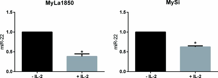
Aberrant activation of Janus kinase-3 (Jak3) and its key down-stream effectors, Signal Transducer and Activator of Transcription-3 (STAT3) and STAT5, is a key feature of malignant transformation in cutaneous T-cell lymphoma (CTCL). However, it remains only partially understood how Jak3/STAT activation promotes lymphomagenesis. Recently, non-coding microRNAs (miRNAs) have been implicated in the pathogenesis of this malignancy. Here, we show that (i) malignant T cells display a decreased expression of a tumor suppressor miRNA, miR-22, when compared to non-malignant T cells, (ii) STAT5 binds the promoter of the miR-22 host gene, and (iii) inhibition of Jak3, STAT3, and STAT5 triggers increased expression of pri-miR-22 and miR-22. Curcumin, a nutrient with anti-Jak3 activity and histone deacetylase inhibitors (HDACi) also trigger increased expression of pri-miR-22 and miR-22. Transfection of malignant T cells with recombinant miR-22 inhibits the expression of validated miR-22 targets including NCoA1, a transcriptional co-activator in others cancers, as well as HDAC6, MAX, MYCBP, PTEN, and CDK2, which have all been implicated in CTCL pathogenesis. In conclusion, we provide the first evidence that de-regulated Jak3/STAT3/STAT5 signalling in CTCL cells represses the expression of the gene encoding miR-22, a novel tumor suppressor miRNA.
Keywords: JAK3; STAT3; STAT5; cutaneous T-cell lymphoma (CTCL); miR-22; mycosis fungoides (MF).
Conflict of interest statement
The authors declare no conflict of interest.
Figures


Similar articles
-
STAT5 induces miR-21 expression in cutaneous T cell lymphoma.Oncotarget. 2016 Jul 19;7(29):45730-45744. doi: 10.18632/oncotarget.10160. Oncotarget. 2016. PMID: 27329723 Free PMC article.
-
Deregulation in STAT signaling is important for cutaneous T-cell lymphoma (CTCL) pathogenesis and cancer progression.Cell Cycle. 2014;13(21):3331-5. doi: 10.4161/15384101.2014.965061. Cell Cycle. 2014. PMID: 25485578 Free PMC article.
-
STAT5-mediated expression of oncogenic miR-155 in cutaneous T-cell lymphoma.Cell Cycle. 2013 Jun 15;12(12):1939-47. doi: 10.4161/cc.24987. Epub 2013 May 15. Cell Cycle. 2013. PMID: 23676217 Free PMC article.
-
miRNA in mycosis fungoides and skin inflammation.APMIS. 2013 Nov;121(11):1017-9. doi: 10.1111/apm.12186. Epub 2013 Sep 30. APMIS. 2013. PMID: 24106870 Review.
-
Regulation of the MIR155 host gene in physiological and pathological processes.Gene. 2013 Dec 10;532(1):1-12. doi: 10.1016/j.gene.2012.12.009. Epub 2012 Dec 14. Gene. 2013. PMID: 23246696 Review.
Cited by
-
Contribution of Non-Coding RNAs to Anticancer Effects of Dietary Polyphenols: Chlorogenic Acid, Curcumin, Epigallocatechin-3-Gallate, Genistein, Quercetin and Resveratrol.Antioxidants (Basel). 2022 Nov 28;11(12):2352. doi: 10.3390/antiox11122352. Antioxidants (Basel). 2022. PMID: 36552560 Free PMC article. Review.
-
Angiogenesis in cutaneous T-cell lymphoma - proteomic approaches.Oncol Lett. 2019 May;17(5):4060-4067. doi: 10.3892/ol.2018.9734. Epub 2018 Nov 19. Oncol Lett. 2019. PMID: 30944599 Free PMC article. Review.
-
Effects of resveratrol, curcumin, berberine and other nutraceuticals on aging, cancer development, cancer stem cells and microRNAs.Aging (Albany NY). 2017 Jun 12;9(6):1477-1536. doi: 10.18632/aging.101250. Aging (Albany NY). 2017. PMID: 28611316 Free PMC article. Review.
-
Enolase 1 stimulates glycolysis to promote chemoresistance in gastric cancer.Oncotarget. 2017 Jul 18;8(29):47691-47708. doi: 10.18632/oncotarget.17868. Oncotarget. 2017. PMID: 28548950 Free PMC article.
-
Plasma miR-155, miR-203, and miR-205 are Biomarkers for Monitoring of Primary Cutaneous T-Cell Lymphomas.Int J Mol Sci. 2017 Oct 15;18(10):2136. doi: 10.3390/ijms18102136. Int J Mol Sci. 2017. PMID: 29036928 Free PMC article.
References
-
- Zhang Q, Nowak I, Vonderheid EC, Rook aH, Kadin ME, Nowell PC, Shaw LM. Activation of Jak/STAT proteins involved in signal transduction pathway mediated by receptor for interleukin 2 in malignant T lymphocytes derived from cutaneous anaplastic large T-cell lymphoma and Sezary syndrome. Proc. Natl. Acad. Sci. U.S.A. 1996;93:9148–53. - PMC - PubMed
-
- Nielsen M, Kaltoft K, Nordahl M, Röpke C, Geisler C, Mustelin T, Dobson P, Svejgaard A, Odum N. Constitutive activation of a slowly migrating isoform of Stat3 in mycosis fungoides: tyrphostin AG490 inhibits Stat3 activation and growth of mycosis fungoides tumor cell lines. Proc. Natl. Acad. Sci. U.S.A. 1997;94:6764–9. - PMC - PubMed
-
- Kopp KLM, Kauczok CS, Lauenborg B, Krejsgaard T, Eriksen KW, Zhang Q, Wasik MA, Geisler C, Ralfkiaer E, Becker JC, Ødum N, Woetmann A. COX-2-dependent PGE(2) acts as a growth factor in mycosis fungoides (MF) Leukemia. 2010;24:1179–85. - PubMed
-
- Zhang Y, Wang Y, Yu R, Huang Y, Su M, Xiao C, Martinka M, Dutz JP, Zhang X, Zheng Z, Zhou Y. Molecular Markers of Early-Stage Mycosis Fungoides. J. Invest. Dermatol. 2012;132:1698–1706. - PubMed
Publication types
MeSH terms
Substances
LinkOut - more resources
Full Text Sources
Other Literature Sources
Medical
Research Materials
Miscellaneous
