

Damage-associated molecular patterns generated in osteoarthritis directly excite murine nociceptive neurons through Toll-like receptor 4
- PMID: 26245312
- PMCID: PMC4626273
- DOI: 10.1002/art.39291
Damage-associated molecular patterns generated in osteoarthritis directly excite murine nociceptive neurons through Toll-like receptor 4
Abstract
Objective: To determine whether selected damage-associated molecular patterns (DAMPs) present in the osteoarthritic (OA) joints of mice excite nociceptors through Toll-like receptor 4 (TLR-4).
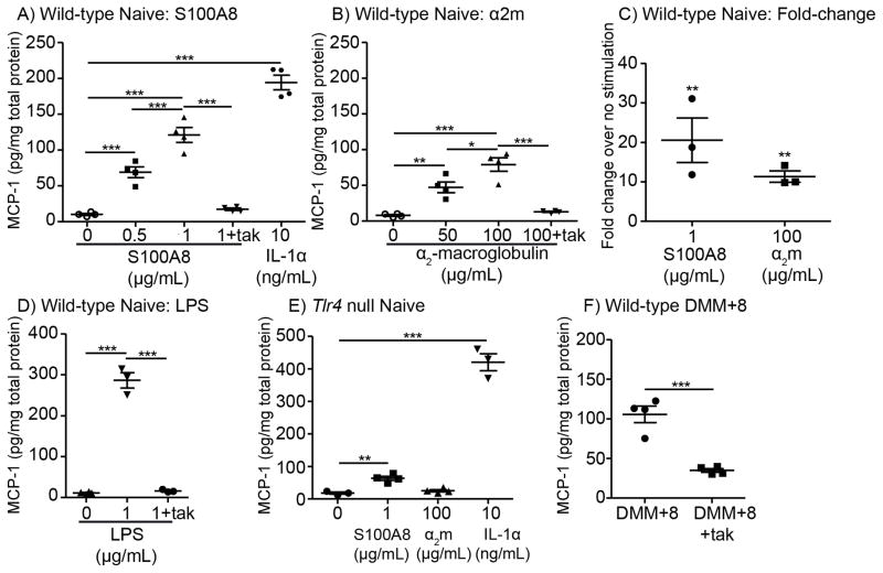
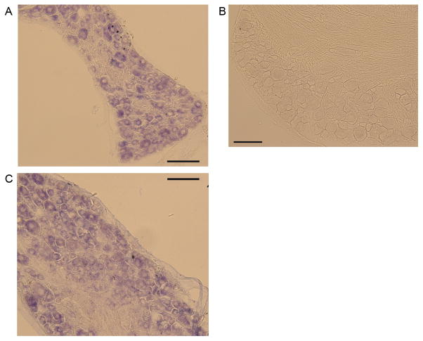
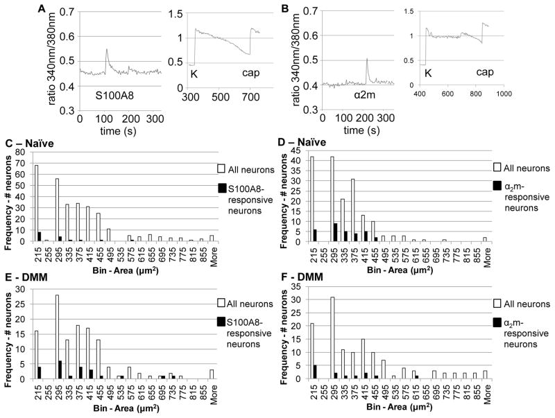
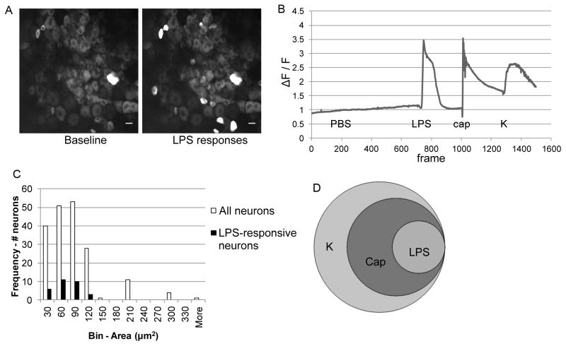
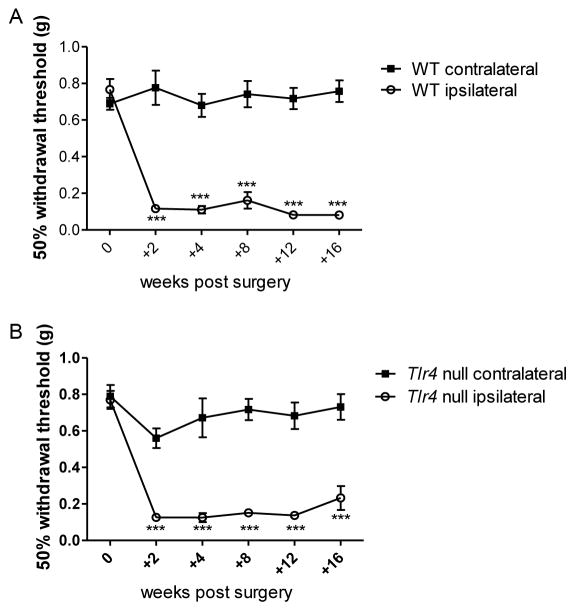
Methods: The ability of S100A8 and α2 -macroglobulin to excite nociceptors was determined by measuring the release of monocyte chemoattractant protein 1 (MCP-1) by cultured dorsal root ganglion (DRG) cells as well as by measuring the intracellular calcium concentration ([Ca(2+) ]i ) in cultured DRG neurons from naive mice or from mice that had undergone surgical destabilization of the medial meniscus (DMM) 8 weeks previously. The role of TLR-4 was assessed using TLR-4(-/-) cells or a TLR-4 inhibitor. The [Ca(2+) ]i in neurons within ex vivo intact DRGs was measured in samples from Pirt-GCaMP3 mice. Neuronal expression of the Tlr4 gene was determined by in situ hybridization. DMM surgery was performed in wild-type and TLR-4(-/-) mice; mechanical allodynia was monitored, and joint damage was assessed histologically after 16 weeks.
Results: DRG neurons from both naive and DMM mice expressed Tlr4. Both S100A8 and α2 -macroglobulin stimulated release of the proalgesic chemokine MCP-1 in DRG cultures, and the neurons rapidly responded to S100A8 and α2 -macroglobulin with increased [Ca(2+) ]i . Blocking TLR-4 inhibited these effects. Neurons within intact DRGs responded to the TLR-4 agonist lipopolysaccharide. In both of the calcium-imaging assays, it was primarily the nociceptor population of neurons that responded to TLR-4 ligands. TLR-4(-/-) mice were not protected from mechanical allodynia or from joint damage associated with DMM.
Conclusion: Our experiments suggest a role of TLR-4 signaling in the excitation of nociceptors by selected DAMPs. Further research is needed to delineate the importance of this pathway in relation to OA pain.
© 2015, American College of Rheumatology.
Figures
Similar articles
-
An emerging role for Toll-like receptors at the neuroimmune interface in osteoarthritis.Semin Immunopathol. 2019 Sep;41(5):583-594. doi: 10.1007/s00281-019-00762-3. Epub 2019 Oct 14. Semin Immunopathol. 2019. PMID: 31612243 Free PMC article. Review.
-
The innate immune response as a mediator of osteoarthritis pain.Osteoarthritis Cartilage. 2020 May;28(5):562-571. doi: 10.1016/j.joca.2019.11.006. Epub 2019 Dec 17. Osteoarthritis Cartilage. 2020. PMID: 31862470 Free PMC article. Review.
-
Notch signaling is activated in knee-innervating dorsal root ganglia in experimental models of osteoarthritis joint pain.Arthritis Res Ther. 2023 Apr 15;25(1):63. doi: 10.1186/s13075-023-03039-1. Arthritis Res Ther. 2023. PMID: 37061736 Free PMC article.
-
Therapeutic effects of an anti-ADAMTS-5 antibody on joint damage and mechanical allodynia in a murine model of osteoarthritis.Osteoarthritis Cartilage. 2016 Feb;24(2):299-306. doi: 10.1016/j.joca.2015.09.005. Epub 2015 Sep 26. Osteoarthritis Cartilage. 2016. PMID: 26410555 Free PMC article.
-
Visualization of Peripheral Neuron Sensitization in a Surgical Mouse Model of Osteoarthritis by In Vivo Calcium Imaging.Arthritis Rheumatol. 2018 Jan;70(1):88-97. doi: 10.1002/art.40342. Epub 2017 Dec 1. Arthritis Rheumatol. 2018. PMID: 28992367 Free PMC article.
Cited by
-
An emerging role for Toll-like receptors at the neuroimmune interface in osteoarthritis.Semin Immunopathol. 2019 Sep;41(5):583-594. doi: 10.1007/s00281-019-00762-3. Epub 2019 Oct 14. Semin Immunopathol. 2019. PMID: 31612243 Free PMC article. Review.
-
The Natural Combination Medicine Traumeel (Tr14) Improves Resolution of Inflammation by Promoting the Biosynthesis of Specialized Pro-Resolving Mediators.Pharmaceuticals (Basel). 2021 Nov 3;14(11):1123. doi: 10.3390/ph14111123. Pharmaceuticals (Basel). 2021. PMID: 34832905 Free PMC article.
-
Role of circular RNAs in osteoarthritis: update on pathogenesis and therapeutics.Mol Genet Genomics. 2023 Jul;298(4):791-801. doi: 10.1007/s00438-023-02021-5. Epub 2023 Apr 22. Mol Genet Genomics. 2023. PMID: 37086279 Review.
-
Decoy peptide targeted to Toll-IL-1R domain inhibits LPS and TLR4-active metabolite morphine-3 glucuronide sensitization of sensory neurons.Sci Rep. 2017 Jun 16;7(1):3741. doi: 10.1038/s41598-017-03447-9. Sci Rep. 2017. PMID: 28623271 Free PMC article.
-
The innate immune response as a mediator of osteoarthritis pain.Osteoarthritis Cartilage. 2020 May;28(5):562-571. doi: 10.1016/j.joca.2019.11.006. Epub 2019 Dec 17. Osteoarthritis Cartilage. 2020. PMID: 31862470 Free PMC article. Review.
References
-
- Cross M, Smith E, Hoy D, Nolte S, Ackerman I, Fransen M, et al. The global burden of hip and knee osteoarthritis: estimates from the global burden of disease 2010 study. Ann Rheum Dis. 2014;73:1323–30. - PubMed
-
- Hochberg MC, Altman RD, April KT, Benkhalti M, Guyatt G, McGowan J, et al. American College of Rheumatology 2012 recommendations for the use of nonpharmacologic and pharmacologic therapies in osteoarthritis of the hand, hip, and knee. Arthritis Care Res (Hoboken) 2012;64:465–74. - PubMed
Publication types
MeSH terms
Substances
Grants and funding
LinkOut - more resources
Full Text Sources
Other Literature Sources
Medical
Research Materials
Miscellaneous
