. 2016 Jun;75(6):1236-45.
doi: 10.1136/annrheumdis-2015-207338. Epub 2015 Aug 5.

Neutrophil microvesicles resolve gout by inhibiting C5a-mediated priming of the inflammasome

Affiliations

Neutrophil microvesicles resolve gout by inhibiting C5a-mediated priming of the inflammasome

Arun Cumpelik et al. Ann Rheum Dis. 2016 Jun.

Abstract

Objectives: Gout is a highly inflammatory but self-limiting joint disease induced by the precipitation of monosodium urate (MSU) crystals. While it is well established that inflammasome activation by MSU mediates acute inflammation, little is known about the mechanism controlling its spontaneous resolution. The aim of this study was to analyse the role of neutrophil-derived microvesicles (PMN-Ecto) in the resolution of acute gout.

Methods: PMN-Ecto were studied in a murine model of MSU-induced peritonitis using C57BL/6, MerTK(-/-) and C5aR(-/-) mice. The peritoneal compartment was assessed for the number of infiltrating neutrophils (PMN), neutrophil microvesicles (PMN-Ecto), cytokines (interleukin-1β, TGFβ) and complement factors (C5a). Human PMN-Ecto were isolated from exudates of patients undergoing an acute gouty attack and functionally tested in vitro.

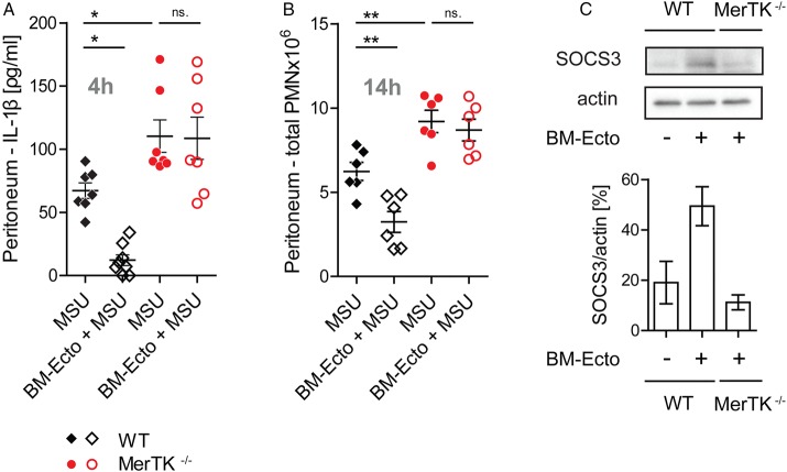
Results: C5a generated after the injection of MSU primed the inflammasome for IL-1β release. Neutrophils infiltrating the peritoneum in response to C5a released phosphatidylserine (PS)-positive PMN-Ecto early on in the course of inflammation. These PMN-Ecto in turn suppressed C5a priming of the inflammasome and consequently inhibited IL-1β release and neutrophil influx. PMN-Ecto-mediated suppression required surface expression of the PS-receptor MerTK and could be reproduced using PS-expressing liposomes. In addition, ectosomes triggered the release of TGFβ independent of MerTK. TGFβ, however, was not sufficient to control acute MSU-driven inflammation in vivo. Finally, PMN-Ecto from joint aspirates of patients with gouty arthritis had similar anti-inflammatory properties.

Conclusions: PMN-Ecto-mediated control of inflammasome-driven inflammation is a compelling concept of autoregulation initiated early on during PMN activation in gout.

Keywords: Cytokines; Gout; Inflammation.


Figures

Figure 1
Figure 1
Characterisation and in vitro properties of BM-Ectosomes. (A) Flow cytometric characterisation of bone marrow-derived ectosomes (BM-Ecto). CFSE-positive events (representing intact vesicles) were analysed for surface expression of Gr-1 and phosphatidylserine (PS; using annexin V) (upper lane, left to right). Controls are (lower lane, left to right): annexin buffer alone, CFSE threshold set on sonicated BM-Ecto previously stained with CFSE, BM-Ecto stained with annexin V and IgG1 isotype in PBS. FSC denotes forward scatter, SSC side scatter, respectively. Numbers indicate % positive BM-Ecto (B) Morphology of BM-Ecto (size bar 100 nm) and (C) monosodium urate (MSU) crystals (size bar 50 µm) as determined by transmission electron microscopy and light microscopy, respectively. (D) In vitro stimulation protocol. B6 peritoneal macrophages were primed with LPS for 10 h and subsequently stimulated with 100 µg/mL MSU for 4 h. BM-Ecto (1×10⁸ BM-Ecto /2×106 macrophages) were given either prior (BM-Ecto+LPS>MSU) or after (LPS>BM-Ecto+MSU) LPS priming as outlined. Alternatively, PS-liposomes or control PC-liposomes were given instead of BM-Ecto. (E and G) IL-1β in cell culture supernatants determined by ELISA. n=4 per group. (F and H) Cell extracts (XT) and supernatants (SN) were analysed for the presence of NALP3, pro-IL-1β, IL-1β and active caspase 1 (p20 and p10) by western blot. Data in A, F and H are representative of three independent experiments. ***p<0.001. Mean±SEM is shown.
Figure 2
Figure 2
Role of C5a in monosodium urate (MSU)-induced inflammation. (A) Inflammasome activation by MSU is dependent on C5a in vitro. Generation of C5a and IL-1β release from C5aR+/+ or C5aR−/− peritoneal macrophages stimulated with 100 µg/mL MSU in the presence of 25% plasma for 14 h in vitro. n=6 per group pooled from two independent experiments. (B and C) BM-Ecto inhibit C5a-mediated inflammasome activation in vitro. (B) BM-Ecto were given to B6 macrophages prior to stimulation with 100 µg/mL MSU in the presence of 25% plasma for 14 h or to (C) macrophages primed with 10 ng/mL recombinant mouse C5a for 10 h and subsequently stimulated with 100 µg/mL MSU for 4 h. IL-1β release, pro-IL-1β and NALP3 expression were analysed as in figure 1. n=6 per group pooled from two independent experiments (ELISA), western blots representative of two independent experiments. (D) Kinetics of MSU-induced peritonitis. B6 mice received an intraperitoneal injection of 3 mg MSU. At the indicated time points thereafter, peritonea were lavaged and infiltrating cells phenotyped by flow cytometry PMN were identified as CD45+, CD11b+, Ly6C+, Ly6G+ cells. Peritoneal concentrations of C5a and IL-1β were determined by ELISA. n=4/time point pooled from two independent experiments. (E) Inflammasome activation is C5a-dependent in vivo. Concentration of IL-1β in the peritoneal lavage fluid of C5aR+/+ and C5aR−/− mice 4 h after injection of 3 mg MSU. n=5 pooled from two independent experiments. (F) Release of BM-Ecto requires the C5aR. 1×10⁷ bone marrow cells were stimulated with 10 ng/mL recombinant mouse C5a for 30 min at 37°C. BM-Ecto were isolated from the supernatant as indicated in Methods. n=5 pooled from two independent experiments. *p<0.05, ***p<0.001. Mean±SEM is shown.
Figure 3
Figure 3
Administration of ectosomes attenuates monosodium urate (MSU)-driven peritoneal inflammation. B6 mice were injected intraperitoneally with 3 mg MSU. Where indicated, mice were preinjected with 2×10⁷ BM-Ecto or PMN-Ecto intraperitoneally 2 h prior to MSU stimulation. Alternatively, mice were preinjected with 75 nM (approximately 2×10⁷) of phosphatidylserine (PS)-liposomes or phosphatidylcholine (PC)-liposomes. Control groups received BM-/PMN-Ecto or PS-/PC-liposome injections intraperitoneally followed by NaCl instead of MSU. (A and C) IL-1β in peritoneal lavage fluid was determined by ELISA 4 h after MSU stimulation. (B and D) The number of infiltrating PMN into the peritoneum 14 h after MSU stimulation was determined as indicated in figure 2D. n=6–8 per group pooled from at least three independent experiments, *p<0.05, **p<0.01, ***p<0.001. Mean±SEM is shown.
Figure 4
Figure 4
Ecto-mediated immunosuppression requires MerTK in vivo. B6/129S (wild type, WT) or MerTK−/− mice were injected intraperitoneally with 3 mg monosodium urate (MSU). Where indicated, mice were preinjected with 2×10⁷ BM-Ecto intraperitoneally 2 h prior to MSU stimulation. (A) IL-1β was determined 4 h after MSU stimulation in the peritoneal lavage fluid. (B) The number of infiltrating PMN into the peritoneum 14 h after MSU stimulation was determined as in figure 2D. n=6–8 per group, pooled from at least three independent experiments. (C) Suppressor of cytokine signalling (SOCS3) expression in peritoneal macrophages of B6/129S (WT) and MerTK−/− mice determined by immunoblot 4 h after intraperitoneal BM-Ecto injection. Expression of SOCS3 in arbitrary units of band intensity normalised to actin. n=2 per group pooled from two independent experiments. *p<0.05, **p<0.05. Mean±SEM is shown. n.s., not significant.
Figure 5
Figure 5
Ecto induce the release of TGF-β independent of MerTK. (A) TGFβ concentration in peritoneal lavage fluids of B6 mice treated as outlined in figure 3 was determined by ELISA. n=6–8 pooled from three independent experiments. (B) Ecto are not the source of TGFβ in vivo. BM-Ecto lysates were assessed for TGFβ content by immunoblot. Recombinant mouse TGFβ was used as control. (C–E) Cellular source of TGFβ. Release of TGFβ by B6 (C) resident peritoneal macrophages or (D) monocytes and neutrophils isolated from monosodium urate (MSU)-inflamed peritonea following treatment with C5a, MSU and/or BM-Ecto in vitro as outlined in figure 2C. (E) Expression of latency associated peptide (LAP) on B6 macrophages, monocytes and neutrophils isolated either 4 h after intraperitoneal injection of BM-Ecto or 4 h after intraperitoneal injection of MSU with BM-Ecto pretreatment. Controls received NaCl intraperitoneally (untreated). (F–H) TGFβ release is independent of MerTK. (F) The release of TGFβ by wild type (WT) and MerTK−/ macrophages treated with BM-Ecto in vitro. n=6/group. TGFβ in peritoneal lavage fluids of (G) B6/129S (WT) and MerTK−/− mice treated with BM-Ecto or (H) B6 mice treated with liposomes as outlined in figures 3 and 4 ,respectively. n=6–8/group. (I and J) Effect of TGFβ in vivo. (I) The effect of neutralising anti-TGFβ1 antibodies on peritoneal PMN influx using 100 µg anti-TGFβ1 injected intraperitoneally 15 min prior to MSU or 15 min prior to BM-Ecto pretreatment. (J) The effect of 1 µg recombinant mouse TGFβ1 injected intraperitoneally instead of BM-Ecto prior to MSU stimulation. *p<0.05, **p<0.01, ***p<0.001. Mean±SEM is shown. n.s., not significant.
Figure 6
Figure 6
PMN-Ecto present in gout exudates in humans have immunosuppressive properties. PMN-Ecto were isolated from joint aspirates of patients undergoing a gout attack. (A) Ecto were isolated from joint aspirates as indicated in methods and characterised and counted by flow cytometry. Annexin V, anti-CD66b and anti-myeloperoxidase (MPO) antibodies identified them to be of neutrophil origin. Counting was performed using microbeads. (B) Transmission electron microscopy of PMN-Ecto. Size bar 1 µm (left) and 100 nm (right). (C) Correlation between the number of infiltrating PMN and PMN-Ecto found in gout exudates. Each dot represents a single patient. (D) Concentration of PMN-Ecto in gout (gouty arthritis, GA) and osteoarthritis (OA) exudates. (E–H) PMN-Ecto isolated from joint aspirates are functional in vitro. (E) Release of TGFβ by human monocyte-derived macrophages (HMDM) treated with Gout-Ecto (1×10⁸ Gout-Ecto/2×106 macrophages). n=7 pooled from two independent experiments. (F) Suppression of IL-1β release by HMDM treated with Gout-Ecto following the in vitro protocol outlined in figure 1D. (G) IL-1β release upon 14 h stimulation of HMDM with 100 µg/mL monosodium urate (MSU) in 25% human plasma (untreated), heat inactivated plasma (heat inactivated) or C5-blocked plasma (αC5). (F and G) n=6 pooled from three independent experiments. (H) Suppression of IL-1β release by HMDM treated with Gout-Ecto and stimulated as outlined in (G). ***p<0.001. Mean±SEM is shown.

Similar articles

Cited by

References

    1. Martinon F, Pétrilli V, Mayor A, et al. . Gout-associated uric acid crystals activate the NALP3 inflammasome. Nature 2006;440:237–41. 10.1038/nature04516 - DOI - PubMed
    1. Rose DM, Sydlaske AD, Agha-Babakhani A, et al. . Transglutaminase 2 limits murine peritoneal acute gout-like inflammation by regulating macrophage clearance of apoptotic neutrophils. Arthritis Rheum 2006;54:3363–71. 10.1002/art.22137 - DOI - PubMed
    1. Steiger S, Harper JL. Neutrophil cannibalism triggers transforming growth factor β1 production and self regulation of neutrophil inflammatory function in monosodium urate monohydrate crystal-induced inflammation in mice. Arthritis Rheum 2013;65:815–23. 10.1002/art.37822 - DOI - PubMed
    1. Huynh ML, Fadok VA, Henson PM. Phosphatidylserine-dependent ingestion of apoptotic cells promotes TGF-beta1 secretion and the resolution of inflammation. J Clin Invest 2002;109:41–50. 10.1172/JCI0211638 - DOI - PMC - PubMed
    1. Yi Z, Li L, Matsushima GK, et al. . A novel role for c-Src and STAT3 in apoptotic cell-mediated MerTK-dependent immunoregulation of dendritic cells. Blood 2009;114:3191–8. 10.1182/blood-2009-03-207522 - DOI - PMC - PubMed

Publication types

MeSH terms