

Critical anatomic region of nasopalatine canal based on tridimensional analysis: cone beam computed tomography
- PMID: 26245884
- PMCID: PMC4526882
- DOI: 10.1038/srep12568
Critical anatomic region of nasopalatine canal based on tridimensional analysis: cone beam computed tomography
Abstract
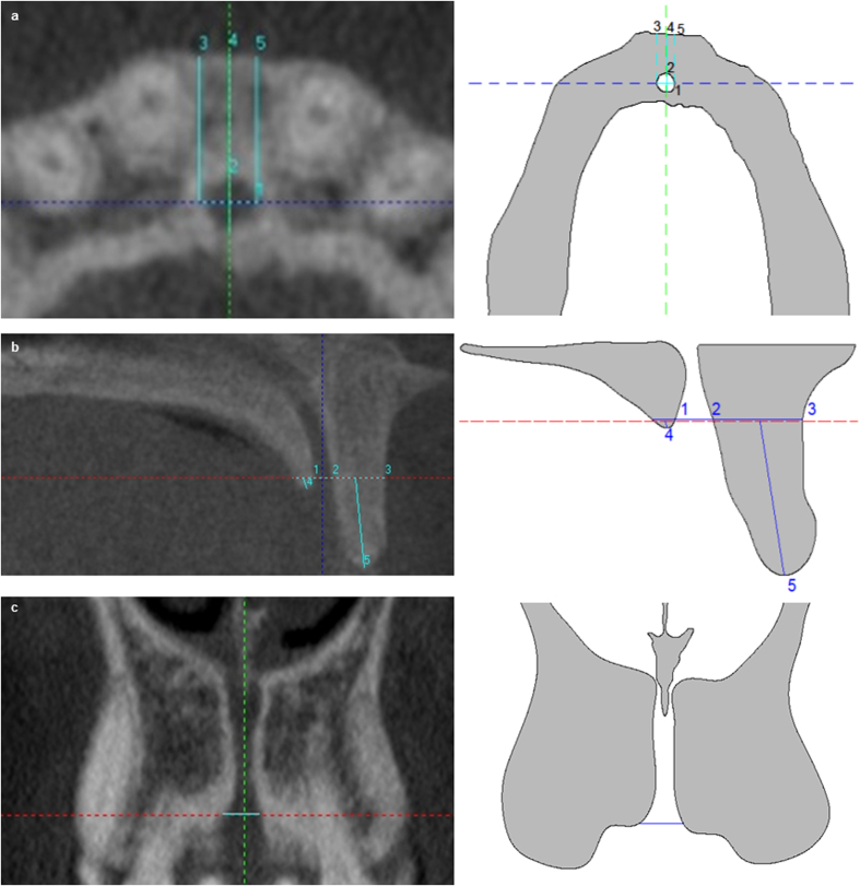
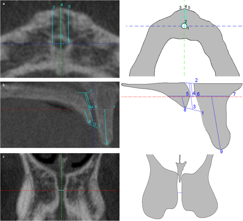
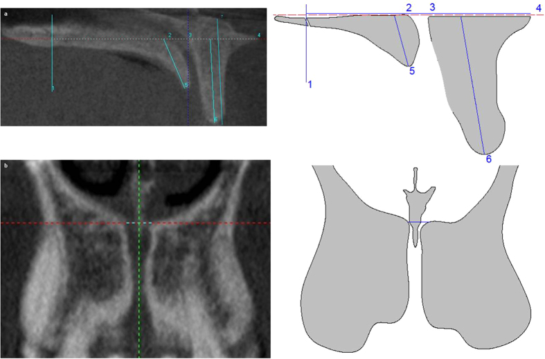
The study aim of this was to define the critical anatomic region of the premaxilla by evaluating dimensions of nasopalatine canal, buccal bone plate (BBP) and palatal bone plate (PBP). 230 CBCTs were selected with both, one or no upper central incisors present (+/+, -/+, -/-) and periodontal condition was evaluated. T-student test, ANOVA, Pearson's correlation and a multivariant-linear regression model (MLRM) were used. Regarding gender, significant differences at level 1 (lower NC) were found for: buccal-palatal, transversal and sagittal NC diameters, and NC length (NCL). Regarding dental status, significant differences were found for: total BBP length (tBL) and PBP width (PW2) at level 2 (NCL midpoint). NCL was correlated with PW2, tBL, and PBP length at level 3 (foramina of Stenson level). An MLRM had a high prediction value for NCL (69.3%). Gender is related to NC dimensions. Dental status has an influence on BBP dimensions, but does not influence on NC and PBP. Periodontal condition should be evaluated for precise premaxillae analysis NC diameters at the three anatomical planes are related to each other, while NCL is related to BBP and PBP lengths. A third of premaxilla is taken up by NC, thus, establishing the critical anatomic region.
Figures
Similar articles
-
Morphometric differences of nasopalatine canal based on 3D classifications: descriptive analysis on CBCT.Surg Radiol Anat. 2015 Sep;37(7):825-33. doi: 10.1007/s00276-015-1470-3. Epub 2015 Mar 29. Surg Radiol Anat. 2015. PMID: 25821035
-
Three-dimensional study of nasopalatine canal morphology: a descriptive retrospective analysis using cone-beam computed tomography.Surg Radiol Anat. 2014 Nov;36(9):895-905. doi: 10.1007/s00276-014-1297-3. Epub 2014 Apr 22. Surg Radiol Anat. 2014. PMID: 24752396
-
Three-Dimensional Analysis of the Anatomical Characteristics and Dimensions of the Nasopalatine Canal Using Cone Beam Computed Tomography.J Maxillofac Oral Surg. 2017 Jun;16(2):197-204. doi: 10.1007/s12663-016-0879-5. Epub 2016 Feb 15. J Maxillofac Oral Surg. 2017. PMID: 28439161 Free PMC article.
-
The Nasopalatine Canal in Adults on Cone Beam Computed Tomograms-A Clinical Study and Review of the Literature.In Vivo. 2015 Jul-Aug;29(4):467-86. In Vivo. 2015. PMID: 26130792 Review.
-
Sex and Population Variations in Nasopalatine Canal Dimensions: A CBCT-Based Systematic Review.Med Sci Monit. 2024 Oct 16;30:e945949. doi: 10.12659/MSM.945949. Med Sci Monit. 2024. PMID: 39410677 Free PMC article.
Cited by
-
Morphological CBCT parameters for an accurate differentiation between nasopalatine duct cyst and the normal nasopalatine canal.Head Face Med. 2024 Sep 28;20(1):54. doi: 10.1186/s13005-024-00458-6. Head Face Med. 2024. PMID: 39342234 Free PMC article.
-
Micro-XCT analysis of anatomical features and dimensions of the incisive canal: implications for dental implant treatment in the anterior maxilla.BMC Oral Health. 2024 Oct 18;24(1):1244. doi: 10.1186/s12903-024-05046-3. BMC Oral Health. 2024. PMID: 39425140 Free PMC article.
-
Evaluation of Morphology and Anatomical Measurement of Nasopalatine Canal Using Cone Beam Computed Tomography.J Dent (Tehran). 2016 Aug;13(4):287-294. J Dent (Tehran). 2016. PMID: 28127321 Free PMC article.
-
Cone beam computerized tomography evaluation of incisive canal and anterior maxillary bone thickness for placement of immediate implants.J Indian Prosthodont Soc. 2018 Oct-Dec;18(4):356-363. doi: 10.4103/jips.jips_167_18. J Indian Prosthodont Soc. 2018. PMID: 30449964 Free PMC article.
References
-
- Williams P. L., Warwick R., Dyson M. & Bannister L. H. Gray’s Anatomy [349] (Churchill Livingstone, 1989).
-
- Rouvière H. & Delmas A. Human anatomy: descriptive, topography and funcional [87] (Masson, 1991).
-
- Liang X. et al. Macro- and micro-anatomical, histological and computed tomography scan characterization of the nasopalatine canal. J. Clin. Periodontol. 36, 598–603 (2009). - PubMed
-
- Buser D., Martin W. & Belser U. C. Optimizing esthetics for implant restorations in the anterior maxilla: anatomic and surgical considerations. Int. J. Oral Maxillofac. Implants. 19, 43–61 (2004). - PubMed
-
- Fernández-Alonso A. et al. Three-dimensional study of nasopalatine canal morphology: a descriptive retrospective analysis using cone-beam computed tomography. Surg. Radiol. Anat. 36, 895–905 (2014). - PubMed
MeSH terms
LinkOut - more resources
Full Text Sources
Other Literature Sources
Research Materials
Miscellaneous
