

Risk factors for poor visual outcome in patients with idiopathic intracranial hypertension
- PMID: 26245929
- PMCID: PMC4553022
- DOI: 10.1212/WNL.0000000000001896
Risk factors for poor visual outcome in patients with idiopathic intracranial hypertension
Abstract
Objectives: Determine potential risk factors for progressive visual field loss in the Idiopathic Intracranial Hypertension Treatment Trial, a randomized placebo-controlled trial of acetazolamide in patients with idiopathic intracranial hypertension and mild visual loss concurrently receiving a low sodium, weight reduction diet.
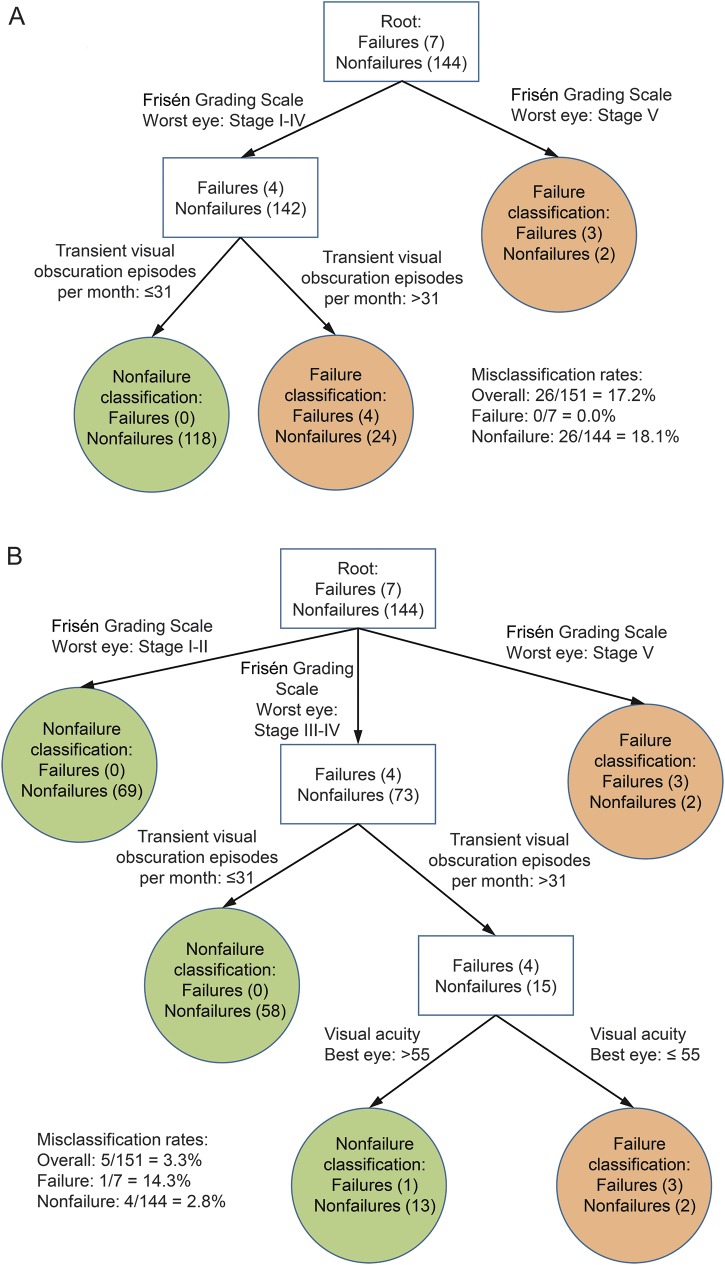
Methods: Logistic regression and classification tree analyses were used to evaluate potential risk factors for protocol-defined treatment failure (>2 dB perimetric mean deviation [PMD] change in patients with baseline PMD -2 to -3.5 dB or >3 dB PMD change with baseline PMD -3.5 to -7 dB).
Results: Seven participants (6 on diet plus placebo) met criteria for treatment failure. The odds ratio for patients with grades III to V papilledema vs those with grades I and II was 8.66 (95% confidence interval [CI] 1.65-∞, p = 0.025). A 1-unit decrease in the number of letters correct on the ETDRS (Early Treatment Diabetic Retinopathy Study) chart at baseline was associated with an increase in the odds of treatment failure by a factor of 1.16 (95% CI 1.04-1.30, p = 0.005). Compared with female participants, the odds ratio for male participants was 26.21 (95% CI 1.61-433.00, p = 0.02). The odds of treatment failure were 10.59 times higher (95% CI 1.63-116.83, p = 0.010) for patients with >30 transient visual obscurations per month vs those with ≤30 per month.
Conclusions: Male patients, those with high-grade papilledema, and those with decreased visual acuity at baseline were more likely to experience treatment failure. All but one of these patients were treated with diet alone. These patients should be monitored closely and be considered for aggressive treatment of their idiopathic intracranial hypertension.
© 2015 American Academy of Neurology.
Figures
References
-
- Smith JL. Whence pseudotumor cerebri? J Clin Neuroophthalmol 1985;5:55–56. - PubMed
-
- Corbett JJ, Savino PJ, Thompson HS, et al. Visual loss in pseudotumor cerebri: follow-up of 57 patients from five to 41 years and a profile of 14 patients with permanent severe visual loss. Arch Neurol 1982;39:461–474. - PubMed
-
- Wall M, George D. Idiopathic intracranial hypertension: a prospective study of 50 patients. Brain 1991;114:155–180. - PubMed
-
- Friedman DI, McDermott MP, Kieburtz K, et al. The idiopathic intracranial hypertension treatment trial: design considerations and methods. J Neuroophthalmol 2014;34:107–117. - PubMed
Publication types
MeSH terms
Substances
Grants and funding
LinkOut - more resources
Full Text Sources
Medical
Miscellaneous