. 2015 Dec;67(12):3135-45.
doi: 10.1002/art.39313.

Release of Active Peptidyl Arginine Deiminases by Neutrophils Can Explain Production of Extracellular Citrullinated Autoantigens in Rheumatoid Arthritis Synovial Fluid

Affiliations

Release of Active Peptidyl Arginine Deiminases by Neutrophils Can Explain Production of Extracellular Citrullinated Autoantigens in Rheumatoid Arthritis Synovial Fluid

Julia Spengler et al. Arthritis Rheumatol. 2015 Dec.

Abstract

Objective: In the majority of patients with rheumatoid arthritis (RA), antibodies specifically recognize citrullinated autoantigens that are generated by peptidylarginine deiminases (PADs). Neutrophils express high levels of PAD and accumulate in the synovial fluid (SF) of RA patients during disease flares. This study was undertaken to test the hypothesis that neutrophil cell death, induced by either NETosis (extrusion of genomic DNA-protein complexes known as neutrophil extracellular traps [NETs]) or necrosis, can contribute to production of autoantigens in the inflamed joint.

Methods: Extracellular DNA was quantified in the SF of patients with RA, patients with osteoarthritis (OA), and patients with psoriatic arthritis (PsA). Release of PAD from neutrophils was investigated by Western blotting, mass spectrometry, immunofluorescence staining, and PAD activity assays. PAD2 and PAD4 protein expression, as well as PAD enzymatic activity, were assessed in the SF of patients with RA and those with OA.

Results: Extracellular DNA was detected at significantly higher levels in RA SF than in OA SF (P < 0.001) or PsA SF (P < 0.05), and its expression levels correlated with neutrophil concentrations and PAD activity in RA SF. Necrotic neutrophils released less soluble extracellular DNA compared to NETotic cells in vitro (P < 0.05). Higher PAD activity was detected in RA SF than in OA SF (P < 0.05). The citrullinated proteins PAD2 and PAD4 were found attached to NETs and also freely diffused in the supernatant. PAD enzymatic activity was detected in supernatants of neutrophils undergoing either NETosis or necrosis.

Conclusion: Release of active PAD isoforms into the SF by neutrophil cell death is a plausible explanation for the generation of extracellular autoantigens in RA.


Figures

Figure 1
Figure 1
Presence of extracellular DNA in the synovial fluid (SF) of patients with rheumatoid arthritis (RA) and correlation with neutrophil cell counts. A, Quantification of extracellular DNA concentrations (conc) in the SF of patients with RA (n = 54), patients with psoriatic arthritis (PsA) (n = 12), and patients with osteoarthritis (OA) (n = 15). Freshly isolated neutrophils from the peripheral blood (PB) of healthy donors (n = 6) were used as a negative control. B, Comparison of free DNA levels in the SF between anti–citrullinated protein antibody (ACPA)–positive and ACPA‐negative RA patients. In A and B, symbols represent individual patients; horizontal bars show the median.  = P < 0.05; ∗∗ = P < 0.01; ∗∗∗ = P < 0.001, by Mann‐Whitney U test. C and D, Assessment of correlations between DNA levels and the neutrophil concentration (C) and macrophage concentration (D) in the SF of 20 RA patients. Correlations were determined by Spearman's rho test.
Figure 2
Figure 2
Detection of neutrophil extracellular traps in the synovial fluid (SF) of patients with rheumatoid arthritis (RA) and attached to the synovial tissue of RA patients. A, SF preparations from 17 RA patients were stained for extracellular DNA (gray) and neutrophil elastase (NE) (green). B, Neutrophil aggregates attached to the synovial tissue of 3 RA patients were detected by confocal laser scanning microscopy. Samples were labeled with antibodies specific for CD15, NE, and citrullinated histone 3 (cit H3) (green) as well as for DNA (gray and blue). Original magnification × 630.
Figure 3
Figure 3
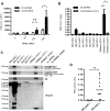
Release of peptidyl arginine deiminases (PADs) into the supernatant during NETosis, and detection of enzymatically active PADs. A, DNA release in peripheral blood neutrophils from healthy donors that were left unstimulated or stimulated with 25 mM phorbol myristate acetate (PMA) was assessed for up to 240 minutes after stimulation. Bars show the mean ± SD of 8 samples per group. B, For isolation of neutrophil extracellular traps, supernatants (SN) of unstimulated (unst.) or stimulated (stim.) neutrophils were collected. Subsequently, the cells were washed 3 times with RPMI medium (W1–W3), and stimulated cells were incubated in the absence or presence of DNase I; unstimulated cells treated with DNase I served as a control. Bars show the mean ± SD DNA concentration (conc) in 7 samples per group. C, Proteins (PAD2, PAD4, and neutrophil elastase [NE]) were precipitated from the same supernatants of unstimulated or stimulated cells as described in B and analyzed by Western blotting. Citrullinated proteins (citr.prot.) were detected using chemical modification and anti–modified citrulline antibody. PAD2 and PAD4 antibodies were tested for cross‐reactivity using human (hu) recombinant PAD4 (rPAD4) (250 ng) and human skeletal muscle tissue lysate (15 μg). One representative blot of 4 independent experiments is shown for each group. D, PAD activity in supernatants of unstimulated and stimulated cells was compared. Symbols represent individual donors (n = 10); bars show the median.  = P < 0.05; ∗∗ = P < 0.01, by Wilcoxon's matched pairs signed rank test. AFU = arbitrary fluorescence units; NS = not significant.
Figure 4
Figure 4
Time course of peptidyl arginine deiminase 2 (PAD2) and PAD4 release during NETosis. Unstimulated (unstim.) peripheral blood neutrophils from healthy donors and neutrophils at 30, 120, or 240 minutes after stimulation were assessed by confocal microscopy, revealing that a proportion of cells enter NETosis after 120 minutes of stimulation. In resting neutrophils, PAD2 is seen only in the cytosol, whereas PAD4 is localized in the nucleus. PAD2 levels drop within 30 minutes after stimulation, and PAD4 is lost from the nuclei of cells undergoing DNA decondensation. No PAD4 signal can be detected after 240 minutes of stimulation; these cells have reached a stage of NETosis at which the nuclear morphology has changed. DNA is shown in blue, PAD2 in green, PAD4 in red, and colabeling with neutrophil elastase (NE) in green. Insets show higher‐magnification views. Original magnification × 400; × 1,200 in insets.
Figure 5
Figure 5
Detection of enzymatically active peptidyl arginine deiminase (PAD) in the synovial fluid (SF) of patients with rheumatoid arthritis (RA). A, Immunoblotting of albumin‐depleted SF samples from 8 RA patients shows presence of neutrophil elastase (NE), PAD4, and PAD2. B, PAD activity in the SF of patients with RA (n = 12) was significantly higher than that in the SF of patients with osteoarthritis (OA) (n = 9). C, PAD activity was observed in the OA SF and RA SF both at physiologic calcium concentrations and at higher calcium concentrations (5 mM). In B and C, symbols represent individual patients; horizontal bars show the median. D, PAD activity is significantly correlated with DNA levels in the SF of RA patients (n = 22). Data were analyzed using Spearman's rho test for correlation.  = P < 0.05; ∗∗ = P < 0.01, by Mann‐Whitney U test.

References

    1. Wright HL, Moots RJ, Edwards SW. The multifactorial role of neutrophils in rheumatoid arthritis. Nat Rev Rheumatol 2014;10:593–601. - PubMed
    1. Geering B, Simon HU. Peculiarities of cell death mechanisms in neutrophils. Cell Death Differ 2011;18:1457–69. - PMC - PubMed
    1. Brinkmann V, Reichard U, Goosmann C, Fauler B, Uhlemann Y, Weiss DS, et al. Neutrophil extracellular traps kill bacteria. Science 2004;303:1532–5. - PubMed
    1. Leon SA, Revach M, Ehrlich GE, Adler R, Petersen V, Shapiro B. DNA in synovial fluid and the circulation of patients with arthritis. Arthritis Rheum 1981;24:1142–50. - PubMed
    1. Khandpur R, Carmona‐Rivera C, Vivekanandan‐Giri A, Gizinski A, Yalavarthi S, Knight JS, et al. NETs are a source of citrullinated autoantigens and stimulate inflammatory responses in rheumatoid arthritis. Sci Transl Med 2013;5:178ra40. - PMC - PubMed

Publication types

MeSH terms