. 2015 Aug 5;35(31):11094-104.
doi: 10.1523/JNEUROSCI.0440-15.2015.

Glutamate Receptors in the Central Nucleus of the Amygdala Mediate Cisplatin-Induced Malaise and Energy Balance Dysregulation through Direct Hindbrain Projections

Affiliations

Glutamate Receptors in the Central Nucleus of the Amygdala Mediate Cisplatin-Induced Malaise and Energy Balance Dysregulation through Direct Hindbrain Projections

Amber L Alhadeff et al. J Neurosci. .

Abstract

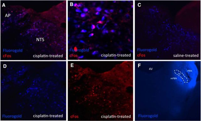
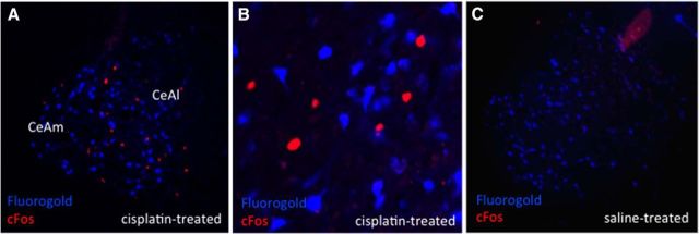
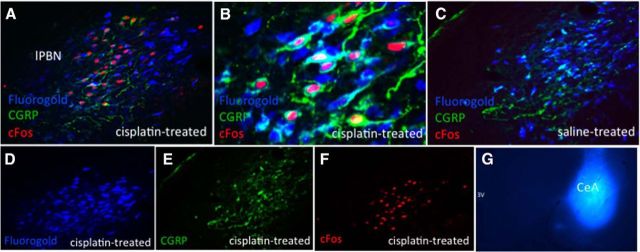
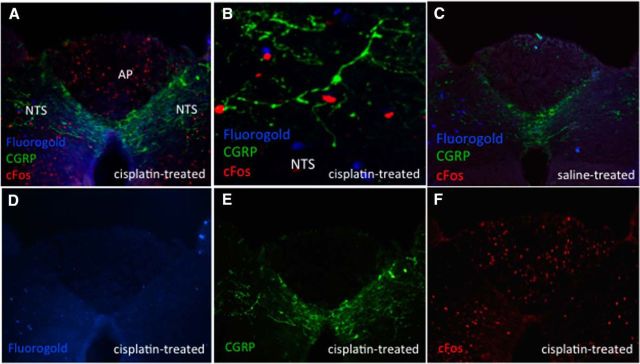
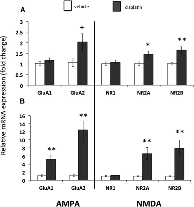
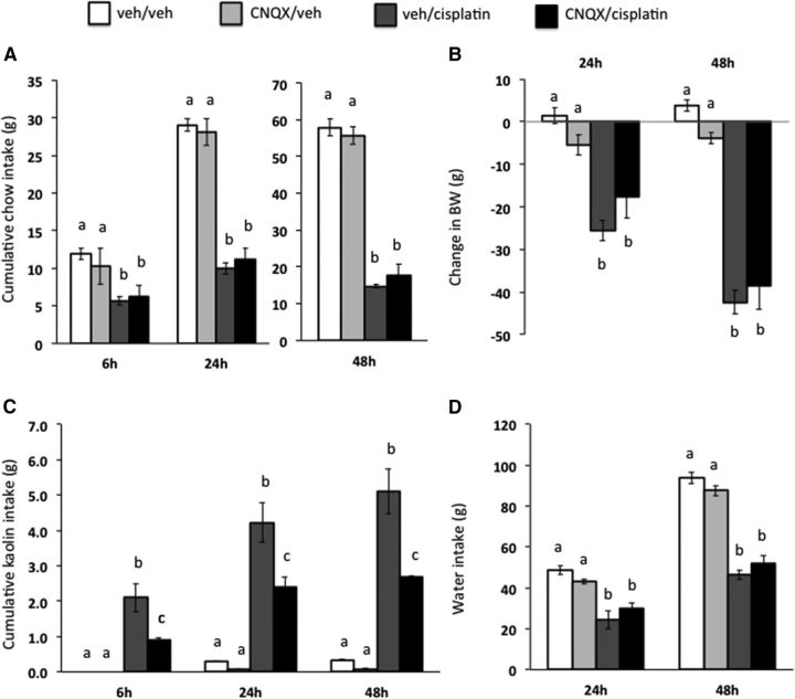
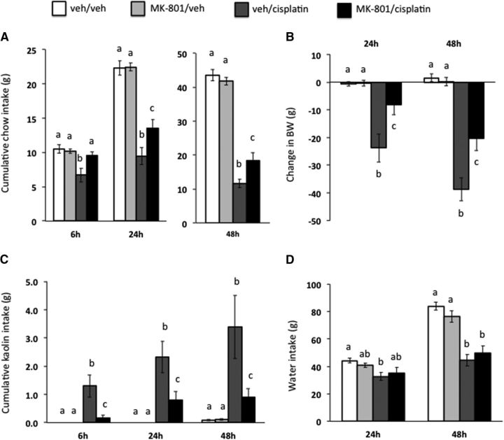
Cisplatin chemotherapy is used commonly to treat a variety of cancers despite severe side effects such as nausea, vomiting, and anorexia that compromise quality of life and limit treatment adherence. The neural mechanisms mediating these side effects remain elusive despite decades of clinical use. Recent data highlight the dorsal vagal complex (DVC), lateral parabrachial nucleus (lPBN), and central nucleus of the amygdala (CeA) as potential sites of action in mediating the side effects of cisplatin. Here, results from immunohistochemical studies in rats identified a population of cisplatin-activated DVC neurons that project to the lPBN and a population of cisplatin-activated lPBN calcitonin gene-related peptide (CGRP, a marker for glutamatergic neurons in the lPBN) neurons that project to the CeA, outlining a neuroanatomical circuit that is activated by cisplatin. CeA gene expressions of AMPA and NMDA glutamate receptor subunits were markedly increased after cisplatin treatment, suggesting that CeA glutamate receptor signaling plays a role in mediating cisplatin side effects. Consistent with gene expression results, behavioral/pharmacological data showed that CeA AMPA/kainate receptor blockade attenuates cisplatin-induced pica (a proxy for nausea/behavioral malaise in nonvomiting laboratory rodents) and that CeA NMDA receptor blockade attenuates cisplatin-induced anorexia and body weight loss in addition to pica, demonstrating that glutamate receptor signaling in the CeA is critical for the energy balance dysregulation caused by cisplatin treatment. Together, these data highlight a novel circuit and CGRP/glutamatergic mechanism through which cisplatin-induced malaise and energy balance dysregulation are mediated.

Significance statement: To treat cancer effectively, patients must follow prescribed chemotherapy treatments without interruption, yet most cancer treatments produce side effects that devastate quality of life (e.g., nausea, vomiting, anorexia, weight loss). Although hundreds of thousands of patients undergo chemotherapies each year, the neural mechanisms mediating their side effects are unknown. The current data outline a neural circuit activated by cisplatin chemotherapy and demonstrate that glutamate signaling in the amygdala, arising from hindbrain projections, is required for the full expression of cisplatin-induced malaise, anorexia, and body weight loss. Together, these data help to characterize the neural circuits and neurotransmitters mediating chemotherapy-induced energy balance dysregulation, which will ultimately provide an opportunity for the development of well tolerated cancer and anti-emetic treatments.

Keywords: amygdala; anorexia; cisplatin; dorsal vagal complex; glutamate; parabrachial nucleus.


Figures

Figure 1.
Figure 1.
A, Representative image of bilateral cannulae tracks and injection site in CeA used for Experiments 4 and 5. B, Timeline of injections in Experiments 4 and 5.
Figure 2.
Figure 2.
DVC cisplatin-activated neurons project directly to the ipsilateral lPBN. Blue immunofluorescence represents FG-expressing neurons and red immunofluorescence represents cisplatin-induced cFos-expressing neurons. A, D, E, Representative DVC images from an lPBN FG- and intraperitoneal cisplatin-treated animal. B, Representative 20× DVC image from an lPBN FG- and intraperitoneal cisplatin-treated animal, C, Representative DVC image from an lPBN FG- and intraperitoneal saline-treated animal. F, Representative lPBN FG injection. n = 4 cisplatin-treated, n = 3 saline-treated. bc, Brachium conjuntivum; mPBN, medial parabrachial nucleus.
Figure 3.
Figure 3.
CeA cisplatin-activated neurons do not project directly to the lPBN. Blue immunofluorescence represents FG-expressing neurons and red immunofluorescence represents cisplatin-induced cFos-expressing neurons. A, Representative CeA image from an lPBN FG and intraperitoneal cisplatin-treated animal. B, Representative 20× CeA image from an lPBN FG- and intraperitoneal cisplatin-treated animal. C, Representative CeA image from an lPBN FG- and intraperitoneal saline-treated animal. n = 5 cisplatin-treated, n = 5 saline-treated. CeAl, Lateral part of CeA; CeAm, medial part of CeA.
Figure 4.
Figure 4.
lPBN cisplatin-activated CGRP/glutamate neurons project directly to the ipsilateral CeA. Blue immunofluorescence represents FG-expressing neurons, red immunofluorescence represents cisplatin-induced cFos-expressing neurons, green immunofluorescence represents CGRP-expressing neurons. A, D, E, F, Representative lPBN images from a CeA FG- and intraperitoneal cisplatin-treated animal. B, Representative 20× lPBN image from a CeA FG- and intraperitoneal cisplatin-treated animal, C, Representative lPBN image from a CeA FG- and intraperitoneal saline-treated animal. G, Representative CeA FG injection. n = 5 cisplatin-treated, n = 4 saline-treated.
Figure 5.
Figure 5.
DVC cisplatin-activated neurons do not project directly to the CeA. Blue immunofluorescence represents FG-expressing neurons, red immunofluorescence represents cisplatin-induced cFos-expressing neurons, green immunofluorescence represents CGRP-expressing neurons. A, D, E, F, Representative DVC image from a CeA FG- and intraperitoneal cisplatin-treated animal. B, Representative 20× NTS image from a CeA FG- and intraperitoneal cisplatin-treated animal. C, Representative DVC image from a CeA FG- and intraperitoneal saline-treated animal. n = 3 cisplatin-treated, n = 4 saline-treated.
Figure 6.
Figure 6.
CeA AMPA and NMDA receptor subunit expressions were increased in cisplatin-treated rats. A, Changes in glutamate receptor subunit expression 6 h after cisplatin (n = 7) or vehicle (n = 5) injection. B, Changes in glutamate receptor subunit expression 24 h after cisplatin (n = 4) or vehicle (n = 5) injection. Data expressed as mean ± SEM. +p = 0.077; *p < 0.05; **p < 0.01 compared with vehicle treatment.
Figure 7.
Figure 7.
CeA AMPA/kainate receptor blockade attenuated cisplatin-induced pica (as measured by kaolin intake, a proxy for nausea/malaise). The selective AMPA/kainate receptor antagonist CNQX significantly attenuated cisplatin-induced kaolin intake (C) and had no effect on cisplatin-induced reduction in chow intake (A), reduction in body weight (B), or reduction in water intake (D). Data are expressed as mean ± SEM, n = 11. Different letters denote significant differences between treatment groups in post hoc comparisons within each time point (p < 0.05).
Figure 8.
Figure 8.
CeA NMDA receptor blockade attenuated cisplatin-induced anorexia, body weight loss, and pica (as measured by kaolin intake, a proxy for nausea/malaise). The selective NMDA receptor antagonist MK-801 significantly attenuated cisplatin-induced reduction in chow intake (A), reduction in body weight (B), and kaolin intake (C) with no effect on cisplatin-induced reduction in water intake (D). Data are expressed as mean ± SEM, n = 12. Different letters denote significant differences between treatment groups in post hoc comparisons within each time point (p < 0.05).

References

    1. Alhadeff AL, Baird JP, Swick JC, Hayes MR, Grill HJ. Glucagon-like Peptide-1 receptor signaling in the lateral parabrachial nucleus contributes to the control of food intake and motivation to feed. Neuropsychopharmacology. 2014;39:2233–2243. - PMC - PubMed
    1. Andrews PL, Horn CC. Signals for nausea and emesis: Implications for models of upper gastrointestinal diseases. Auton Neurosci. 2006;125:100–115. doi: 10.1016/j.autneu.2006.01.008. - DOI - PMC - PubMed
    1. Andrews PL, Sanger GJ. Nausea and the quest for the perfect anti-emetic. Eur J Pharmacol. 2014;722:108–121. doi: 10.1016/j.ejphar.2013.09.072. - DOI - PubMed
    1. Bence KK, Delibegovic M, Xue B, Gorgun CZ, Hotamisligil GS, Neel BG, Kahn BB. Neuronal PTP1B regulates body weight, adiposity and leptin action. Nat Med. 2006;12:917–924. doi: 10.1038/nm1435. - DOI - PubMed
    1. Buse JB, Rosenstock J, Sesti G, Schmidt WE, Montanya E, Brett JH, Zychma M, Blonde L, LEAD-6 Study Group Liraglutide once a day versus exenatide twice a day for type 2 diabetes: a 26-week randomised, parallel-group, multinational, open-label trial (LEAD-6) Lancet. 2009;374:39–47. doi: 10.1016/S0140-6736(09)60659-0. - DOI - PubMed

Publication types

MeSH terms