. 2015 Aug 6:13:58.
doi: 10.1186/s12915-015-0167-8.

Role of electrostatic interactions for ligand recognition and specificity of peptide transporters

Affiliations

Role of electrostatic interactions for ligand recognition and specificity of peptide transporters

Rajendra Boggavarapu et al. BMC Biol. .

Abstract

Background: Peptide transporters are membrane proteins that mediate the cellular uptake of di- and tripeptides, and of peptidomimetic drugs such as β-lactam antibiotics, antiviral drugs and antineoplastic agents. In spite of their high physiological and pharmaceutical importance, the molecular recognition by these transporters of the amino acid side chains of short peptides and thus the mechanisms for substrate binding and specificity are far from being understood.

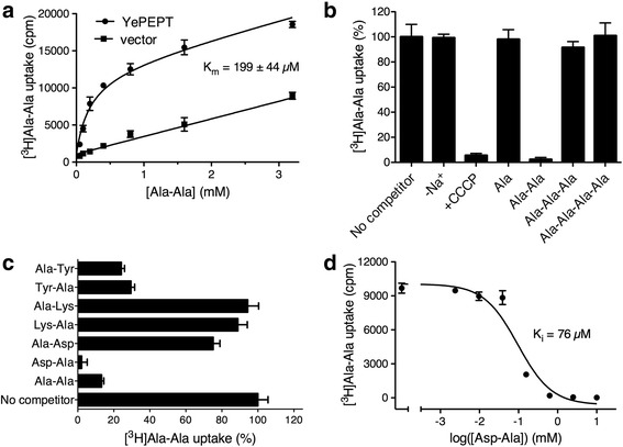
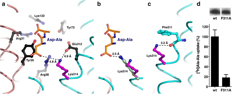
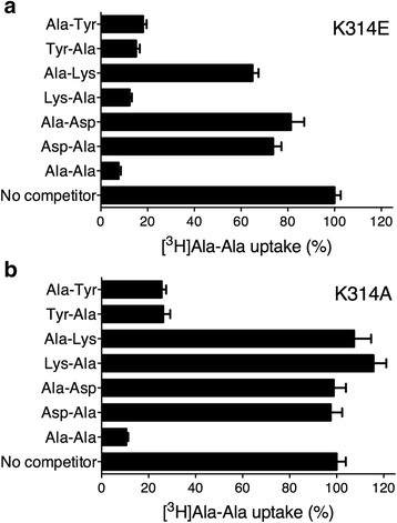
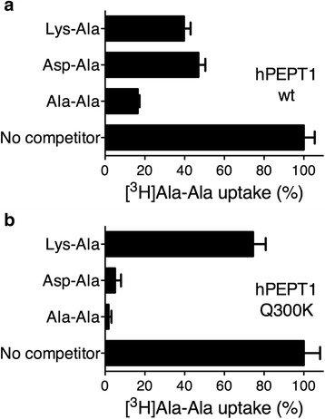
Results: The X-ray crystal structure of the peptide transporter YePEPT from the bacterium Yersinia enterocolitica together with functional studies have unveiled the molecular bases for recognition, binding and specificity of dipeptides with a charged amino acid residue at the N-terminal position. In wild-type YePEPT, the significant specificity for the dipeptides Asp-Ala and Glu-Ala is defined by electrostatic interaction between the in the structure identified positively charged Lys314 and the negatively charged amino acid side chain of these dipeptides. Mutagenesis of Lys314 into the negatively charged residue Glu allowed tuning of the substrate specificity of YePEPT for the positively charged dipeptide Lys-Ala. Importantly, molecular insights acquired from the prokaryotic peptide transporter YePEPT combined with mutagenesis and functional uptake studies with human PEPT1 expressed in Xenopus oocytes also allowed tuning of human PEPT1's substrate specificity, thus improving our understanding of substrate recognition and specificity of this physiologically and pharmaceutically important peptide transporter.

Conclusion: This study provides the molecular bases for recognition, binding and specificity of peptide transporters for dipeptides with a charged amino acid residue at the N-terminal position.


Figures

Fig. 1
Fig. 1
Functional characterization of YePEPT. a Kinetics of YePEPT-mediated [3H]Ala-Ala uptake in E. coli cells. Uptake of the radioligand in E. coli cells transformed with the YePEPT construct (YePEPT) and the empty vector (vector; control) is shown. The determined Km is indicated. Error bars represent SEM from triplicates. One representative experiment from three similar independent experiments is shown. b Co-transport ion and substrate chain length dependence of uptake: Na+ dependence was assessed by replacing Na+ with choline (−Na+); and H+ dependence by addition of the proton-ionophore carbonyl cyanide 3-chlorophenylhydrazone (CCCP). Chain length dependence was assessed with L-Ala and the corresponding di-, tri- and tetrapeptides as competitors (10 mM final concentration). c Substrate specificity of YePEPT by competition assay (2.5 mM final concentration). Error bars in (b) and (c) represent SEM from at least three independent experiments, each in triplicate. d Ki determination of YePEPT for Asp-Ala. The determined Ki is indicated (95 % confidence intervals: 46–126 μM). Error bars represent SEM from triplicates. One representative experiment from three similar independent experiments is shown
Fig. 2
Fig. 2
Overall structure of YePEPT. Structure of YePEPT viewed in the plane of the membrane (top) and from the cytosol (bottom). The N- and C-terminal six-helix bundles are colored in red and blue, respectively. The two helices HA and HB connecting the two bundles are colored in gold. The N- and C-termini are labeled. In the top view (bottom) the transmembrane helices H1–H12, and HA and HB are labeled. Parts of the loops connecting H6 and HA, and H8 and H9 that could not be traced are indicated by broken lines
Fig. 3
Fig. 3
Substrate-binding pocket of YePEPT. Top view from the cytosol (a) and view from the membrane plane (d) of the substrate-binding pocket of YePEPT. The areas marked by the black boxes in (a) and (d) are displayed at higher magnification in (b) and (c), and (e) and (f), respectively. Amino acid residues of YePEPT potentially involved in alafosfalin (in yellow; (b) and (e)) and dipeptide backbone binding (Ala-Phe dipeptide in green; panels (c) and (f)) are labeled, colored in black and conserved in YePEPT, GkPOTE310Q and PepTSt (see Additional file 6: Table S2 for more details and groups of conserved amino acid residues involved in alafosfalin and Ala-Phe dipeptide backbone binding). The indicated amino acid residues of YePEPT were identified by superposition of the YePEPT structure with the ligand-bound peptide transporter structures 4D2C [8] and 4IKZ [9] (PDB ID codes). The distances between the nitrogen atom of Lys314 and the carbon atoms of the N-terminally located methyl groups of alafosfalin (e) and Ala-Phe (f) are marked, and indicate available space between ligands and Lys314; for example, for accommodation of longer side chains. The N- and C-terminal six-helix bundles are colored in salmon and cyan, respectively, and helices HA and HB in black. YePEPT polypeptide chains in (a) and (d) that could not be traced are indicated by broken lines
Fig. 4
Fig. 4
Hypothetical model of Asp-Ala bound YePEPT and stabilizing effect of Phe311. a YePEPT structure with a virtually bound Asp-Ala dipeptide (view from the membrane plane): this dipeptide was built by keeping the dipeptide backbone of Ala-Phe (Fig. 3c,f) fixed and mutating the side chains of Ala-Phe into Asp-Ala in Pymol [23]. The rotamer of Asp in Asp-Ala does not introduce any clashes with the YePEPT structure. The distances between the nitrogen atom of Lys314 and the closest oxygen atoms of the carboxyl groups of Asp-Ala (residue at R1 position) and Glu312 are indicated. b The rotamer of Lys314 found in the crystal structure (magenta) and of the rotamer with the shortest distance to the closest oxygen atom of the carboxyl group of Asp-Ala (residue at R1 position) is shown (pale green). c In the YePEPT crystal structure, the interactions between Lys314 and the stabilizing residue Phe311 consist of a hydrogen bond and a pi-cation interaction between the ε-amino group of Lys314, and the carbonyl and phenyl groups of Phe311, respectively. Amino acid residues of YePEPT potentially involved in dipeptide backbone binding are labeled and colored in black (similar to Fig. 3). The N- and C-terminal six-helix bundles are colored in salmon and cyan, respectively. d Mutation of the Lys314 stabilizing Phe311 residue into Ala dramatically reduces the transport function of YePEPT. Expression levels in E. coli of wild-type (wt) and YePEPTF311A (F311A) used for the uptake experiments were comparable. Error bars in (d) represent SEM from two independent experiments, each in triplicate
Fig. 5
Fig. 5
Substrate specificity of K314E and K314A YePEPT mutants. [3H]Ala-Ala uptake competition experiments performed with E. coli cells expressing (a) K314E and (b) K314A mutants of YePEPT. Error bars represent SEM from four independent experiments, each in triplicate
Fig. 6
Fig. 6
Comparison of substrate specificities of human PEPT1 and Q300K mutant. [3H]Ala-Ala uptake competition experiments were performed with Xenopus laevis oocytes expressing (a) wild-type human PEPT1 (hPEPT1-wt) and (b) the Q300K mutant (hPEPT1-Q300K). Error bars represent SEM from two independent experiments, each containing 12 oocytes

References

    1. Paulsen IT, Skurray RA. The POT family of transport proteins. Trends Biochem Sci. 1994;19:404. doi: 10.1016/0968-0004(94)90087-6. - DOI - PubMed
    1. Daniel H, Spanier B, Kottra G, Weitz D. From bacteria to man: archaic proton-dependent peptide transporters at work. Physiology (Bethesda) 2006;21:93–102. doi: 10.1152/physiol.00054.2005. - DOI - PubMed
    1. Daniel H, Kottra G. The proton oligopeptide cotransporter family SLC15 in physiology and pharmacology. Pflugers Arch. 2004;447:610–8. doi: 10.1007/s00424-003-1101-4. - DOI - PubMed
    1. Rubio-Aliaga I, Daniel H. Mammalian peptide transporters as targets for drug delivery. Trends Pharmacol Sci. 2002;23:434–40. doi: 10.1016/S0165-6147(02)02072-2. - DOI - PubMed
    1. Brandsch M. Drug transport via the intestinal peptide transporter PepT1. Curr Opin Pharmacol. 2013;13:881–7. doi: 10.1016/j.coph.2013.08.004. - DOI - PubMed

Publication types