. 2015 Oct;93(4):88.
doi: 10.1095/biolreprod.115.131227. Epub 2015 Aug 5.

Geography of follicle formation in the embryonic mouse ovary impacts activation pattern during the first wave of folliculogenesis

Affiliations

Geography of follicle formation in the embryonic mouse ovary impacts activation pattern during the first wave of folliculogenesis

Marília H Cordeiro et al. Biol Reprod. 2015 Oct.

Abstract

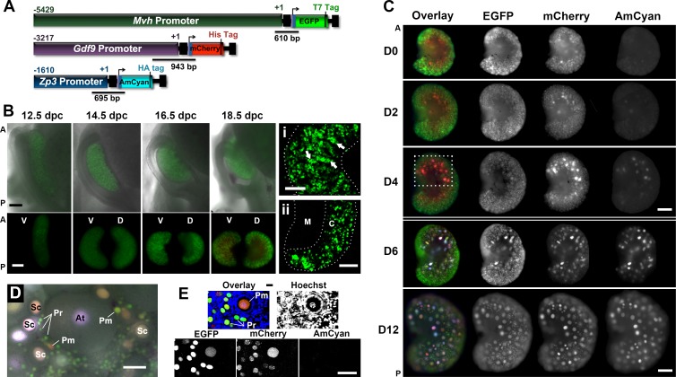
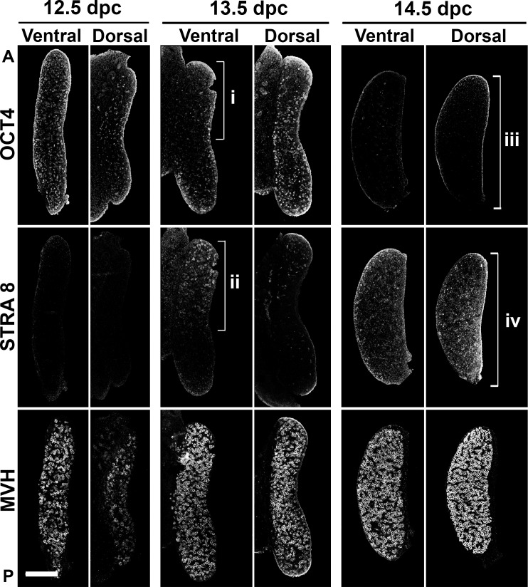
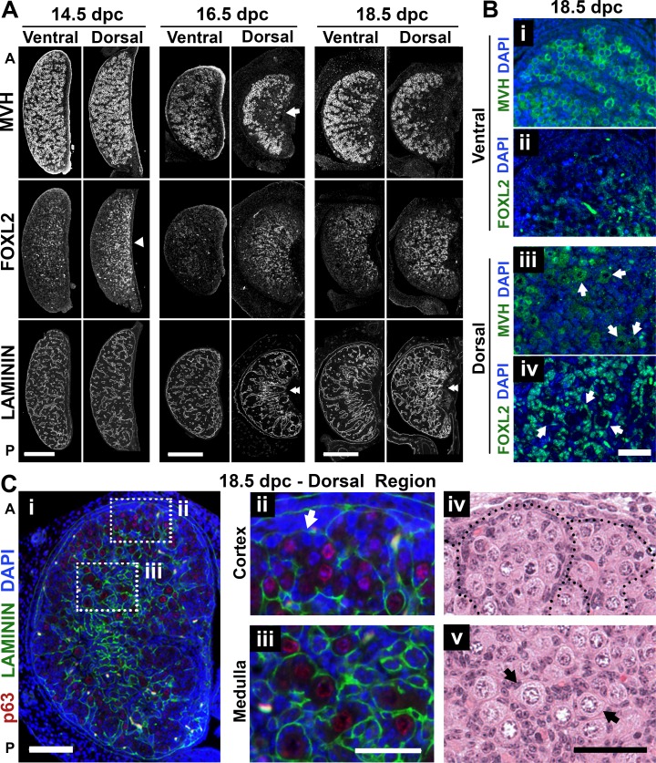
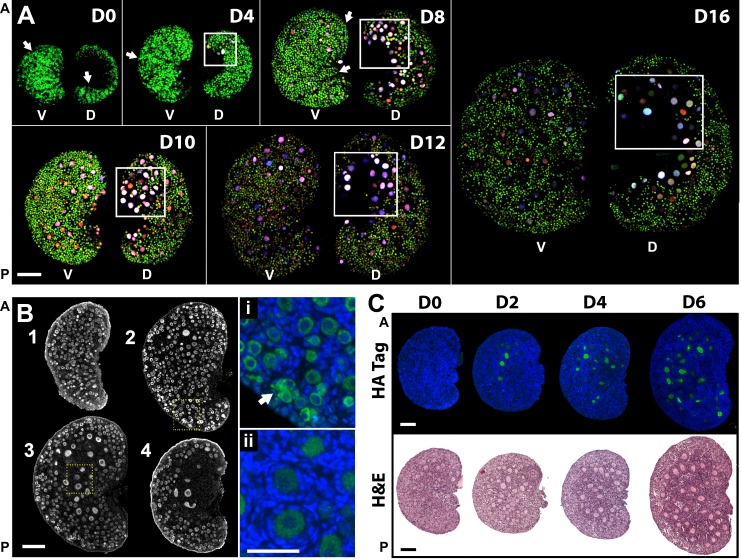
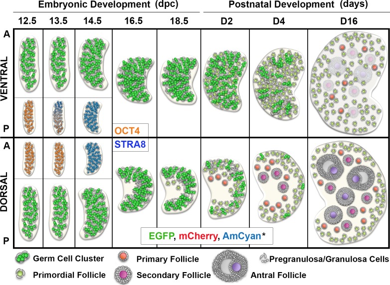
During embryonic development, mouse female germ cells enter meiosis in an anterior-to-posterior wave believed to be driven by retinoic acid. It has been proposed that ovarian follicle formation and activation follow the same general wave of meiotic progression; however, the precise anatomic specification of these processes has not been delineated. Here, we created a mouse line using Mvh, Gdf9, and Zp3 promoters to drive distinct temporal expression of three fluorescent proteins in the oocytes and to identify where the first follicle cohort develops. The fluorescent profile revealed that the first growing follicles consistently appeared in a specific region of the ovary, the anterior-dorsal region, which led us to analyze if meiotic onset occurred earlier in the dorsal ovarian region. Surprisingly, in addition to the anterior-to-posterior wave, we observed an early meiotic entry in the ventral region of the ovary. This additional anatomic stratification of meiosis contrasts with the localization of the initial follicle formation and activation in the dorsal region of the ovary. Therefore, our study suggests that the specification of cortical and medullar areas in the ventral and dorsal regions on the ovary, rather than the onset of meiosis, impacts where the first follicle activation event occurs.

Keywords: follicle activation; follicle formation; meiosis; ovarian geography; ovary.


Figures

FIG. 1
FIG. 1
Expression of the fluorescent proteins changed according to the developmental stage of the germ cell. A) Map of the linear constructs used to generate the triple-transgenic line and design of the genotyping primers. B) Expression of reporter proteins during embryonic development (epifluorescence). Top) Anatomic changes of the ovary and reproductive tract. Bottom) Ventral (V) and dorsal (D) regions of the same ovary, rotated horizontally. A, anterior; P, posterior. Bar = 200 μm. Detail of the cord-like structures (i; arrows highlight germ cell cluster) and specification of a medullar region (ii) in 18.5-dpc ovaries (confocal maximum projections) are shown. M, medulla; C, cortex. Bar in i and ii = 100 μm. C) Fluorescent protein expression in the dorsal ovarian region of different age mouse (epifluorescence). Dashed box highlights the ovarian area where follicles first grow. Different imaging settings were used at D0 to D4 and D6 to D12. A higher EGFP and lower AmCyan and mCherry exposure times were used on D6 and D12 to avoid saturation of the images. Bar = 200 μm. D) Representative follicles in the surface of the ventral region in a 16-day-old mouse ovary (epifluorescence). Follicle stage was determined based on the follicular morphology in the bright-field image. Pr, primordial; Pm, primary; Sc, secondary; At, antral. Bar = 50 μm. E) Small follicles in a 16-day-old mouse ovary (confocal). Primordial follicles (Pr) only express EGFP and mCherry at various levels. DNA probe Hoechst 33342 was used to determine follicle class. Bar = 50 μm.
FIG. 2
FIG. 2
Timing of meiosis entry differed between ventral and dorsal regions of the mouse ovary. Meiosis onset was visualized using immunofluorescence assay in serial sections with OCT4, STRA8, and MVH antibodies, allowing detection of germ cells before, during, and independent of the meiotic entry, respectively. Early onset of meiosis in the anterior-ventral region at 13.5 dpc as visualized by the depletion of OCT4 (i) and expression of STRA8 (ii) is shown. At 14.5 dpc, the dorsal region of the ovary also showed evidence of meiosis (iii and iv). Note the uniform distribution of germ cells in both regions of the gonad, visualized with MVH immunofluorescence assay. 12.5 dpc, n = 11; 13.5 dpc, n = 15; 14.5 dpc, n = 9. A, anterior; P, posterior. Bar = 200 μm.
FIG. 3
FIG. 3
Timing of individual follicle formation differed between ventral and dorsal regions of the mouse ovary. A) Differences between dorsal and ventral ovarian regions identified using immunofluorescence assay with MVH (germ cell), FOXL2 (pregranulosa), and laminin (basal lamina) antibodies in serial sections. Arrow highlights the specification of the medulla at 16.5 dpc. Arrowhead shows higher density of FOXL2-expressing cells in the dorsal region even before specification of the medulla. Double-arrowheads highlight dorsal-ventral differences in laminin organization. 14.5 dpc, n = 9; 16.5 dpc, n = 7; 18.5 dpc, n = 9. Bar = 200 μm. B) Individual follicle formation at 18.5 dpc was restricted to the dorsal region of the ovary, visualized using MVH (green; i and iii) and FOXL2 (green; ii and iv). Arrows represent individual primordial follicles. The images in i/ii and iii/iv are adjacent sections separated by 5 μm. Bar = 50 μm. C) Spatial differences in development between the center and the periphery of the dorsal region at 18.5 dpc (i) along with details of the germ cell clusters at the periphery (ii and iv) and individual follicles in the center (iii and v). Arrows highlight the presence of individual follicles. Red, p63 (oocyte nucleus); green, laminin; blue, DAPI. A, anterior; P, posterior. Bar = 100 μm (i) and 50 μm (ii–v).
FIG. 4
FIG. 4
Differential follicle development in ventral and dorsal ovarian regions is conserved throughout the prepubertal period. A) Maximum projection of confocal z-stack images revealed different oocyte populations on the surface of the two ovarian regions. Boxes highlight regions with elevated number of growing follicles (anterior-dorsal). Arrows exemplify follicle distribution resembling cord-like structures. The same imaging settings were used except Day 0 and Day 4, which required lower laser power for EGFP detection. Ventral (V) and dorsal (D) regions of the same ovary, rotated horizontally, are shown. Scale bars are approximated because images represent three-dimensional projections; n = 5 for each time point. Bar = ∼200 μm. B) Geographic differences in follicle development in the same ovary. Immunofluorescence assay with MVH antibody in a 4-day-old mouse ovary in serial sections 45 μm apart (n = 6). Arrow shows region where ovarian nest remains. Highlighted areas are expanded on i and ii. 1, ventral; 4, dorsal. Green, MVH; blue, DAPI. Bar = 100 μm and 50 μm (i and ii). C) Gradual expansion of the ovarian region containing growing follicles; n = 6–10 for each represented time point. Green, HA tag; blue, DAPI; H&E, hematoxylin and eosin staining. A, anterior; P, posterior. Bar = 100 μm.
FIG. 5
FIG. 5
Schematics of developmental events in the ventral and dorsal ovarian regions during the embryonic period and first wave of follicle activation. Early onset of meiosis in the ventral region is represented in the insert (OCT4 and STRA8). First primordial follicles were observed at 18.5 dpc at the interface between cortex and medulla in the anterior-dorsal region of the ovary, the area where the first cohort of growing follicles appear during the first week of postnatal development. In contrast, during this same period, germ cells in the ventral region of the ovary are still breaking the nest and forming individual follicles. The area containing growing follicles expands gradually during the postnatal period, occupying the whole medullar area of the dorsal region of the ovary. Altogether, our data indicated that the first wave of follicle activation follows a predictable anatomic pattern resulting from regional differences in timing of follicle formation. *Color resulting from the overlay of the three fluorescent proteins is represented. A, anterior; P, posterior.

Comment in

References

    1. Baker TG. Radiosensitivity of mammalian oocytes with particular reference to the human female. Am J Obstet Gynecol. 1971;110:746–761. - PubMed
    1. Kerr JB, Myers M, Anderson RA. The dynamics of the primordial follicle reserve. Reproduction. 2013;146:R205–R215. - PubMed
    1. Menke DB, Koubova J, Page DC. Sexual differentiation of germ cells in XX mouse gonads occurs in an anterior-to-posterior wave. Dev Biol. 2003;262:303–312. - PubMed
    1. Bullejos M, Koopman P. Germ cells enter meiosis in a rostro-caudal wave during development of the mouse ovary. Mol Reprod Dev. 2004;68:422–428. - PubMed
    1. Bowles J, Knight D, Smith C, Wilhelm D, Richman J, Mamiya S, Yashiro K, Chawengsaksophak K, Wilson MJ, Rossant J, Hamada H, Koopman P. Retinoid signaling determines germ cell fate in mice. Science. 2006;312:596–600. - PubMed

Publication types

MeSH terms