. 2015 Sep 8;6(26):22641-52.
doi: 10.18632/oncotarget.4010.

Role of PAR-4 in ovarian cancer

Affiliations

Role of PAR-4 in ovarian cancer

Sonia Meynier et al. Oncotarget. .

Abstract

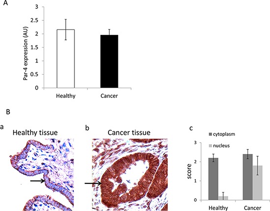
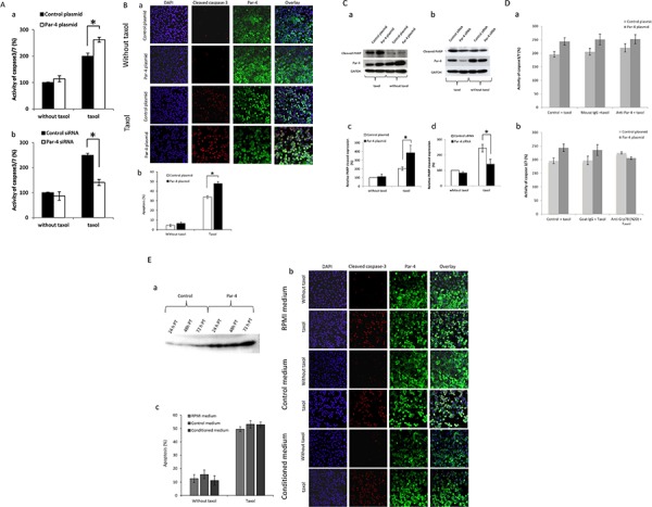
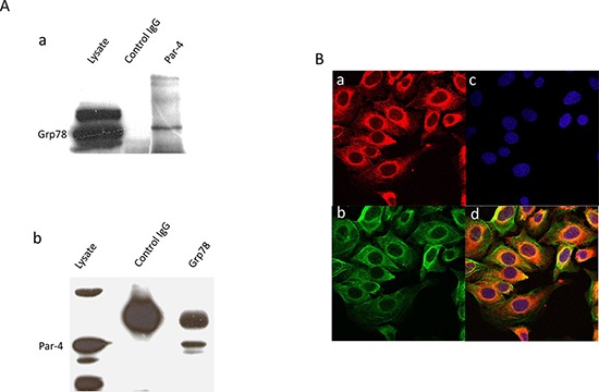
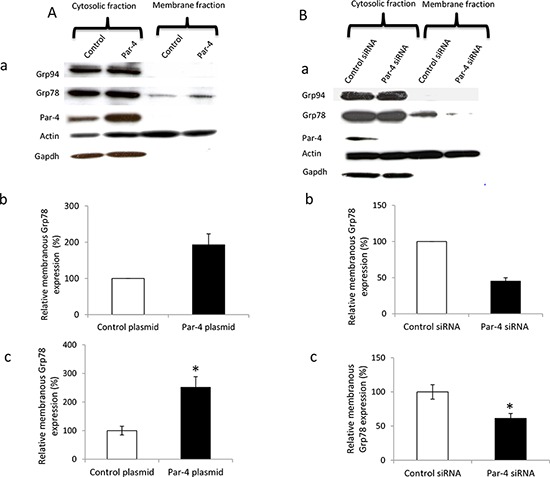
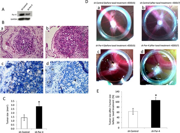
Prostate apoptosis response-4 (PAR-4) is considered as a tumour suppressor due to its ability to selectively induce cell apoptosis in most cancer cells. However little is known about the role of PAR-4 in ovarian cancer. In this study, we investigated for the first time the role of PAR-4 in ovarian carcinogenesis. We showed that PAR-4 mRNA level is not significantly different between healthy and cancer ovarian cells. Immunohistochemistry on ovarian tissue showed that ovarian cancer cells are positive for PAR-4 nuclear and cytoplasmic staining whereas ovarian healthy cells are negative for PAR-4 nuclear staining. We then studied the role of PAR-4 in cell apoptosis. We determined that PAR-4 induces cell apoptosis in response to stimuli, in vitro, but is also involved in the relocation of GRP78 from endoplasmic reticulum to the cell surface of ovarian cancer cell line (SKOV-3 cells). In ovo, PAR-4 decreases ovarian tumour development and increases the response to taxol treatment. These observations suggest that PAR-4 is a very interesting therapeutic target against ovarian carcinogenesis.

Keywords: GRP78; PAR-4; apoptosis; ovarian cancer.


Conflict of interest statement

CONFLICTS OF INTEREST

There is no conflicts of interest in our study.

Figures

Figure 1
Figure 1. Presence of PAR-4 in healthy and cancer tissues
A. Presence of PAR-4 mRNA in healthy and cancer tissues. qPCR analysis of PAR-4 mRNA expression in healthy and cancer cells. Two housekeeping genes were used: GAPDH and Cyclophilin A. B. Expression of PAR-4 in healthy and cancer tissues. Immunohistochemistry of healthy (a) and high grade serous ovarian cancer (b) tissues with control normal rabbit IgG and anti-PAR-4 antibodies (R-334). The magnification used is ×200. Arrows indicate epithelial cells. (c) Score of staining intensity established by two experts for the cytoplasm and the nucleus PAR-4 levels.
Figure 2
Figure 2. Effect of PAR-4 levels on cell apoptosis
A. Analysis of caspase 3/7 activity. SKOV-3 cells are transfected with control or PAR-4 expressing plasmid (a) and with control or PAR-4 siRNA (b) and treated or not with 100 nM of taxol. After 24 hours, luminescent assay is performed to measure caspase3/7 activity. Quantitative results are presented in Figures a and b. B. Apoptosis scoring of active caspase-3. SKOV-3 cells are transfected with control or PAR-4 expressing plasmid. 24 hours after, SKOV-3 cells are seeded and treated or not with 100 nM of taxol for 24 hours. SKOV-3 cells are washed, fixed and then, incubated with anti-PAR-4 antibodies (R-334) and active caspase-3 antibodies. Then, cells are incubated for 1 hour with Alexa Fluor-488 donkey anti-rabbit IgG or Alexa Fluor-568 donkey anti-mouse IgG. The nucleus is stained with DAPI. SKOV-3 cells are analysed with LSM 510 META. The magnification used is ×200 (a). The percentage of apoptotic cells is calculated as follows: Apoptosis % = number of active caspase 3 positive cells/number of nuclei (b). C. Analysis of cleaved PARP by Western Blot. SKOV-3 cells are transfected with control or PAR-4 expressing plasmid (a) and with control or PAR-4 siRNA (b) and treated or not with 100 nM of taxol. After 48 hours, cell extracts are collected. Western Blot is performed and probed with anti-cleaved-PARP and anti-GAPDH antibodies. Bands of western blot are revealed by an ECL method, scanned and quantified by the Kodak 1D image analysis software. The cleaved-PARP band intensity is normalized to GAPDH and is expressed in percentage in function of control plasmid without taxol (c, d). D. Analysis of caspase3/7 activity with inhibition of PAR-4. SKOV-3 cells transfected with control or PAR-4 expressing plasmid are treated or not with 100 nM of taxol and 2.5 ug/ml of anti-PAR-4 antibodies (a) or anti-GRP78 antibodies (N20) (b) or IgG control for 24 hours. After 24 hours, caspase3/7 activity is examined by luminescent assay 24 post-treatment with antibodies. Quantitative results are presented in Figures a and b. E. Active caspase-3 staining in SKOV-3 cells treated or not with conditioned media. SKOV-3 cells are transfected with control or PAR-4 expressing plasmids. The culture supernatant is collected at 24, 48 h and 72 h post transfection (PT), and centrifuged at 14 000 rpm for 10 min. The supernatant is then aliquoted and frozen until use. Western blot is performed to evaluate the presence of secreted PAR-4 in medium (a). SKOV-3 cells are transfected with control or PAR-4 and GRP78 expressing plasmids. The efficiency of transfection is confirmed by western blot (data not shown). 24 hours after transfection, SKOV-3 cells are seeded in Lab-tek chamber. 24 hours after, SKOV-3 cells are treated or not with 100 nM of taxol and with 72 h PT conditioned medium for 24 hours. SKOV-3 cells are washed, fixed and then, incubated with anti-PAR-4 antibodies (R-334) and active caspase-3 antibodies. Then, cells are incubated for 1 hour with Alexa Fluor-488 donkey anti-rabbit IgG or Alexa Fluor-568 donkey anti-mouse IgG. The nucleus is stained with DAPI. SKOV-3 cells are analysed with LSM 510 META. The magnification used is ×200 (b). The percentage of apoptotic cells is calculated as follows: Apoptosis % = number of active caspase 3 positive cells/number of nuclei (c).
Figure 3
Figure 3. Colocation of PAR-4 and GRP78
A. Co-immunoprecipitation. a. Western blot analysis of SKOV-3 cell lysate immunoprecipitated with anti-PAR-4 antibodies and blotted with GRP78 (Gl-19) antibodies. b. Western blot analysis of SKOV-3 cell lysate immunoprecipitated with anti-GRP78 (Gl-19) antibodies and blotted with anti-PAR-4 antibodies. B. Immunofluorescence. Immunofluorescence assay of SKOV-3 cells is performed with anti-GRP78 (N20) (a), and anti-PAR-4 antibodies (b). The nucleus is stained with Dapi (c). The cells are analysed with LSM 510 META. The magnification used is ×400. In the merged images (d), the colocation is shown in yellow.
Figure 4
Figure 4. Effect of PAR-4 expression on GRP78 relocation in ovarian cancer cell surface
A. Effect of PAR-4 overexpression on GRP78 relocation. a. SKOV-3 cells are transfected with control or PAR-4 expressing plasmid. Western blot is done and probed with anti-GRP78, anti-PAR-4, anti-Actin, as loading control, and anti-GAPDH, as cytosol control, antibodies. b. Bands of western blot are revealed by an ECL method, scanned and quantified by the Kodak 1D image analysis software. The GRP78 band intensity is normalized to Actin and is expressed in percentage in function of control plasmid. c. Transfected SKOV-3 cells are seeded at 20 000 cells/well in 96-well plate. After 48 hours, cells are incubated with anti-GRP78 antibodies and then with HRP conjugated goat anti-rabbit IgG antibody. The results are expressed as the relative ration of membranous GRP78 over total GRP78 (*p < 0.05, Student t-test). B. Effect of PAR-4 downregulation on GRP78 relocation. a. SKOV-3 cells are transfected with control or PAR-4 siRNA. Western blot is done and probed with anti GRP78, anti-PAR-4, Actin and GAPDH antibodies. b. Bands of western blot are revealed by an ECL method, scanned and quantified by the Kodak 1D image analysis software. The GRP78 band intensity is normalized to Actin and is expressed in percentage in function of control plasmid. c. Transfected SKOV-3 cells are seeded at 20 000 cells/well in 96-well plate. After 48 hours, cells are incubated with anti-GRP78 antibodies and then with HRP conjugated goat anti-rabbit IgG antibody. The results are expressed as the relative ration of membranous GRP78 over total GRP78 (*p < 0.05, Student t-test).
Figure 5
Figure 5. Influence of PAR-4 on the efficiency of taxol on tumour development
A. Western blot analysis of PAR-4 levels after transfection of SKOV-3 cells with shControl or shPAR-4. B. HE staining of shControl tumour (a) and shPAR-4 tumour (b) tissues. The magnification used is ×200. Numbers 1 indicate tumours and numbers 2 show the chicken membrane. PAR-4 staining of shControl tumour (c) and shPAR-4 (d) tissues. The brown colour represents the PAR-4 expression. The magnification used is ×400. C. Evaluation of the tumour size before taxol treatment on EDD 15 (n = 10 for shControl and n = 14 for shPAR-4) using ImageJ software. D. On EDD8, shControl SKOV-3 cells (a) or shPAR-4 SKOV-3 cells (c) suspension are placed into the silicon O-ring for 4 days to allow tumour growth. On EDD15, shControl (a) or shPAR-4 tumours (c) are treated topically with 25 μl of 25 μM of taxol for 2 days. On EDD17, tumour size of shControl (b) and shPAR-4 (d) is monitored using a Wild Heerbrugg M3Z microscope at 10× magnification with a Lumenera INFINITY2–1 CDD camera with Infinity Capture Software. E. Evaluation of the tumour size after taxol treatment (n = 10 for shControl and n = 14 for shPAR-4) using ImageJ software. The size of tumours on EDD 17 is measured and the average is done.

References

    1. Ferlay J, Shin HR, Bray F, Forman D, Mathers C, Parkin DM. Estimates of worldwide burden of cancer in 2008: GLOBOCAN 2008. Int J Cancer. 127:2893–2917. - PubMed
    1. Clark TG, Stewart ME, Altman DG, Gabra H, Smyth JF. A prognostic model for ovarian cancer. Br J Cancer. 2001;85:944–952. - PMC - PubMed
    1. Sankaranarayanan R, Ferlay J. Worldwide burden of gynaecological cancer: the size of the problem. Best Pract Res Clin Obstet Gynaecol. 2006;20:207–225. - PubMed
    1. Irby RB, Kline CL. Par-4 as a potential target for cancer therapy. Expert Opin Ther Targets. 2013;17:77–87. - PubMed
    1. El-Guendy N, Zhao Y, Gurumurthy S, Burikhanov R, Rangnekar VM. Identification of a unique core domain of par-4 sufficient for selective apoptosis induction in cancer cells. Mol Cell Biol. 2003;23:5516–5525. - PMC - PubMed

Publication types

MeSH terms