. 2015 Aug 21;6(24):20404-18.
doi: 10.18632/oncotarget.4057.

Upregulated long non-coding RNA AFAP1-AS1 expression is associated with progression and poor prognosis of nasopharyngeal carcinoma

Affiliations

Upregulated long non-coding RNA AFAP1-AS1 expression is associated with progression and poor prognosis of nasopharyngeal carcinoma

Hao Bo et al. Oncotarget. .

Abstract

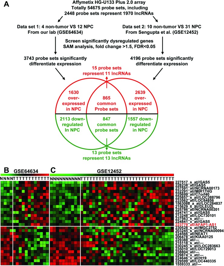
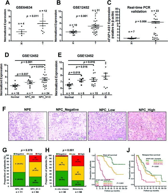
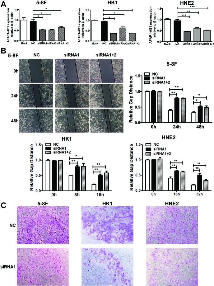
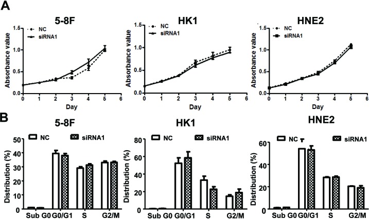
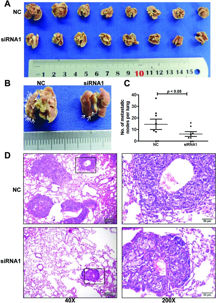
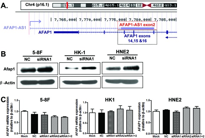
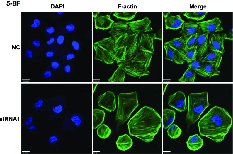
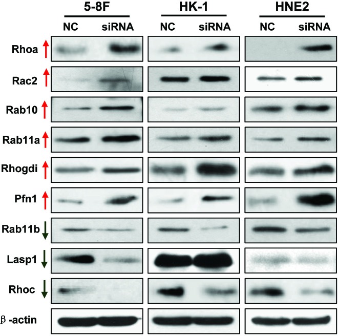
Altered expression of long noncoding RNAs (lncRNAs) associated with human carcinogenesis. We performed a cDNA microarray analysis of lncRNA expression in 12 cases of nasopharyngeal carcinoma (NPC) and 4 non-tumor nasopharyngeal epitheliums. One lncRNA, actin filament associated protein 1 antisense RNA1 (AFAP1-AS1), was identified and selected for further study. AFAP1-AS1 expression was upregulated in NPC and associated with NPC metastasis and poor prognosis. In vitro experiments demonstrated that AFAP1-AS1 knockdown significantly inhibited the NPC cell migration and invasive capability. AFAP1-AS1 knockdown also increased AFAP1 protein expression. Proteomic and bioinformatics analyses suggested that AFAP1-AS1 affected the expression of several small GTPase family members and molecules in the actin cytokeratin signaling pathway. AFAP1-AS1 promoted cancer cell metastasis via regulation of actin filament integrity. AFAP1-AS1 might be a potential novel marker that can predict cancer patient prognosis and as a potential therapeutic target for NPC.

Keywords: AFAP1 antisense RNA1 (AFAP1-AS1); long non-coding RNA (LncRNA); metastasis; nasopharyngeal carcinoma (NPC); prognosis.


Conflict of interest statement

CONFLICTS OF INTEREST STATEMENT

The authors declare that there are no conflicts of interest in this work.

Figures

Figure 1
Figure 1. Dysregulated lncRNA expression analysis using two independent NPC cohorts and cDNA microarray analysis
A. Schematic overview of the workflow used to identify and validate dysregulated lncRNAs in two NPC microarray data cohorts. B. Heatmap of 28 dysregulated probe sets representing 24 lncRNAs mined from our own NPC dataset (GSE64634). C. Heatmap of 28 dysregulated probe sets representing 24 lncRNAs mined from the GSE12452 data set.
Figure 2
Figure 2. Upregulated AFAP1-AS1 was associated with tumor metastasis and poor prognosis
A. AFAP1-AS expression, as measured by Affymetrix Microarray, was upregulated in NPC biopsies (T, n = 12) when compared with non-tumor NPE tissues (N, n = 4). B. Upregulated AFAP1-AS expression was confirmed in NPC biopsies (T, n = 31) compared with non-tumor NPE tissues (N, n = 10). C. AFAP1-AS expression in non-tumor NPE tissues (N, n = 7) and NPC biopsies (T, n = 23) was validated in another cohort of NPC samples using qRT-PCR. D. AFAP1-AS1 expression was associated with lymph node metastasis in the GSE12452 dataset (normal: non-tumor NPE; NPC_N0: NPC biopsies without lymph node metastasis; N1/2: NPC biopsies with lymph node metastasis). E. AFAP1-AS1 expression was associated with clinical disease stages in the GSE12452 dataset (normal: non-tumor NPE; I, II or III: NPC biopsies with clinical stage I, II or III disease). F. AFAP1-AS1 expression measured by in situ hybridization in paraffin embedded non-tumor NPE (N) and NPC biopsies (T). Representative cases of non-tumor NPE (N) and NPC biopsies with negative (n = 34), low (n = 55) or high (n = 23) AFAP1-AS1 staining are shown. G. Proportion of NPC patients with AFAP1-AS1 expression with lymph node metastasis (N0, n = 11; N1/2, n = 97, p = 0.078). H. Proportion of NPC patients with AFAP1-AS1 expression and in situ relapse (n = 30) or distant metastasis (n = 58, p = 0.001). I. and J. Overall survival and relapse-free survival analysis of patients with negative, low and high AFAP1-AS1 staining using a Kaplan-Meier curve, p < 0.001. T, tumor; N, non-tumor tissues.
Figure 3
Figure 3. AFAP1-AS1 knockdown suppressed tumor cell migration and invasion in vitro
A. siRNA knockdown of AFAP1-AS1 expression. siRNA1, siRNA2 and siRNA1+2 dramatically suppressed AFAP1-AS1 expression at the RNA level when compared with Mock and the scrambled control-siRNA (NC) in 5-8F, HK1 and HNE2 cells by qRT-PCR. B. AFAP1-AS1 knockdown inhibited 5-8F, HK1 and HNE2 cell migration. Cells were grown and transfected with AFAP1-AS1 siRNA or scramble siRNA for 24 h and then subjected to the wound healing assay at 0, 24 and 48 h (upper left panel). The data are summarized as the width ratio of migratory inhibition. Control siRNA treated cells efficiently migrated into the gap, while AFAP1-AS1 siRNA treated cells uniformly displayed significantly impaired wound closure (upper right panel and lower panel). C. AFAP1-AS1 knockdown inhibited tumor cell invasion as measured by Transwell Matrigel penetration assay. 5-8F, HK1 and HNE2 cells were grown and transfected with AFAP1-AS1 siRNA, or control siRNA for 36 h and then subjected to a Matrigel invasion assay. The graph summarizes the data from three independent experiments. *p < 0.05, **p < 0.01, ***p< 0.001.
Figure 4
Figure 4. AFAP1-AS1 knockdown didn't affect cell viability cell cycle distribution and apoptosis in vitro
A. Cell viability assays using 5-8F, HK1 and HNE2 cells. Cells were grown and transfected with AFAP1-AS1 siRNA or control siRNA and then subjected to cell viability MTT assays. AFAP1-AS1 knockdown had no effect on cancer cell viability. B. Cell cycle distribution and apoptosis detection. 5-8F, HK1 and HNE2 cells were grown and transfected with AFAP1-AS1 or control siRNA for 36 h and then subjected to flow cytometry cell cycle and apoptosis assays. Relative to the scrambled control-siRNA-transfected cells, the AFAP1-AS1 knockdown had no significant effect on cell cycle distribution or apoptosis.
Figure 5
Figure 5. AFAP1-AS1 knockdown inhibited NPC 5-8F cell metastasis in vivo
A. Bright-field images of mouse lungs taken 10 weeks after injection of 1 × 10⁶ AFAP1-AS1 siRNA or negative control treated 5-8F cells into the tail vein. B. Representative images of visible nodules on the mouse lung surface. Arrows indicate clusters of tumor cells that have colonized the lung. C. Numbers of visible lung metastases in nude mice. The data are presented as the means ± SEM (each data point represents a different mouse; n = 8 mice per group). D. Representative images of AFAP1-AS1 siRNA expressing 5-8F cells after metastasis to lung tissue. H&amp;E-stained sections of lung tissues after the mice received injections of AFAP1-AS1 siRNA expressing 5-8F cells. Rectangular boxes indicate clusters of micro metastatic cells in the lung. Images were acquired at 40X and 200X. Scale bars = 200 µm and 50 µm.
Figure 6
Figure 6. AFAP1-AS1 knockdown upregulated the expression of AFAP1 protein
A. Alignment of AFAP1-AS1 with the protein-coding AFAP1 gene on chromosome 4p16.1. Three exons (14, 15 and 16) of AFAP1 complemented the second exon of AFAP1-AS1. B. AFAP1-AS1 knockdown upregulated AFAP1 protein levels in 5-8F, HK1 and HNE2 cells when compared to the negative control (scrambled control-siRNA). C. AFAP1-AS1 knockdown did not affect AFAP1 mRNA levels in 5-8F, HK1 or HNE2 cells when compared to the negative control (scrambled control-siRNA).
Figure 7
Figure 7. AFAP1-AS1 knockdown induced loss of stress filament integrity in cancer cells
Immunofluorescence staining of 5-8F cells after AFAP-AS1 knockdown. Forty-eight hours after the plating of cells onto fibronectin-coated glass coverslips, cells were fixed and stained for F-actin with phalloidin. A clear deficiency in stress fiber formation was observed in AFAP-AS1 knockdown cells. Images were acquired at 400X. Scale bar = 20 µm.
Figure 8
Figure 8. Validation of differentially expressed proteins identified by proteomics
Differentially expressed proteins were confirmed by using Western blotting in 5-8F, HK1 and HNE2 cancer cell lines transfected with the scrambled control-siRNA or AFAP1-AS1 siRNA. The results were consistent with the proteomic analysis.

References

    1. Wei WI, Sham JS. Nasopharyngeal carcinoma. Lancet. 2005;365:2041–2054. - PubMed
    1. Chang ET, Adami HO. The enigmatic epidemiology of nasopharyngeal carcinoma. Cancer epidemiology, biomarkers &amp; prevention : a publication of the American Association for Cancer Research, cosponsored by the American Society of Preventive Oncology. 2006;15:1765–1777. - PubMed
    1. Lo KW, Chung GT, To KF. Deciphering the molecular genetic basis of NPC through molecular, cytogenetic, and epigenetic approaches. Semin Cancer Biol. 2012;22:79–86. - PubMed
    1. Xiong W, Zeng ZY, Xia JH, Xia K, Shen SR, Li XL, Hu DX, Tan C, Xiang JJ, Zhou J, Deng H, Fan SQ, Li WF, Wang R, Zhou M, Zhu SG, et al. A susceptibility locus at chromosome 3p21 linked to familial nasopharyngeal carcinoma. Cancer Research. 2004;64:1972–1974. - PubMed
    1. Zeng Z, Huang H, Huang L, Sun M, Yan Q, Song Y, Wei F, Bo H, Gong Z, Zeng Y, Li Q, Zhang W, Li X, Xiang B, Li X, Li Y, et al. Regulation network and expression profiles of Epstein-Barr virus-encoded microRNAs and their potential target host genes in nasopharyngeal carcinomas. Science China Life sciences. 2014;57:315–326. - PubMed

Publication types

Substances