. 2015 Oct 15;24(20):5901-14.
doi: 10.1093/hmg/ddv315. Epub 2015 Aug 5.

Endogenous DUX4 expression in FSHD myotubes is sufficient to cause cell death and disrupts RNA splicing and cell migration pathways

Affiliations

Endogenous DUX4 expression in FSHD myotubes is sufficient to cause cell death and disrupts RNA splicing and cell migration pathways

Amanda M Rickard et al. Hum Mol Genet. .

Abstract

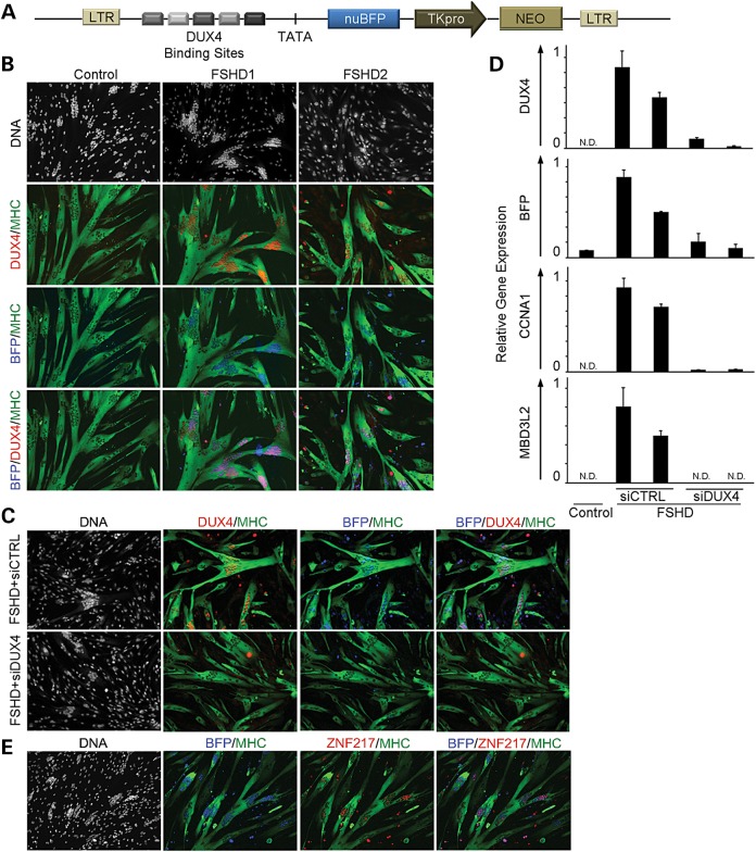
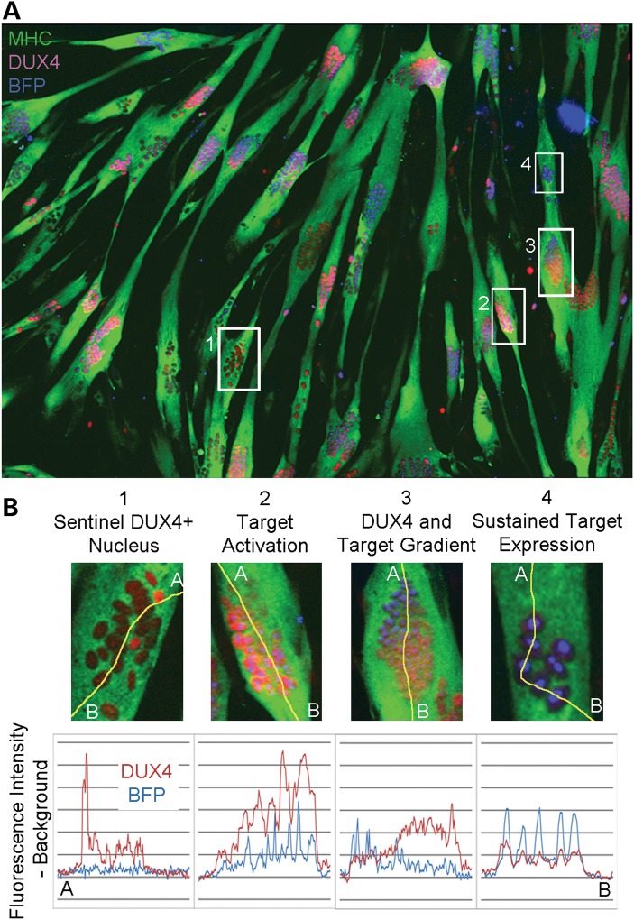
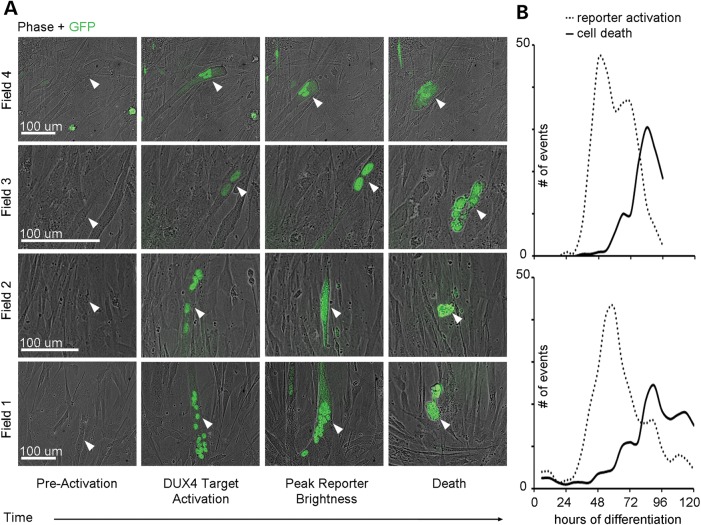
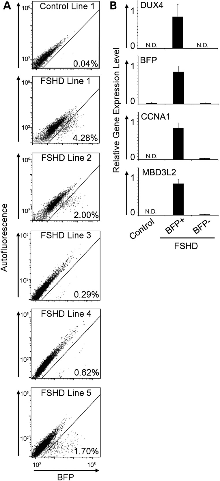
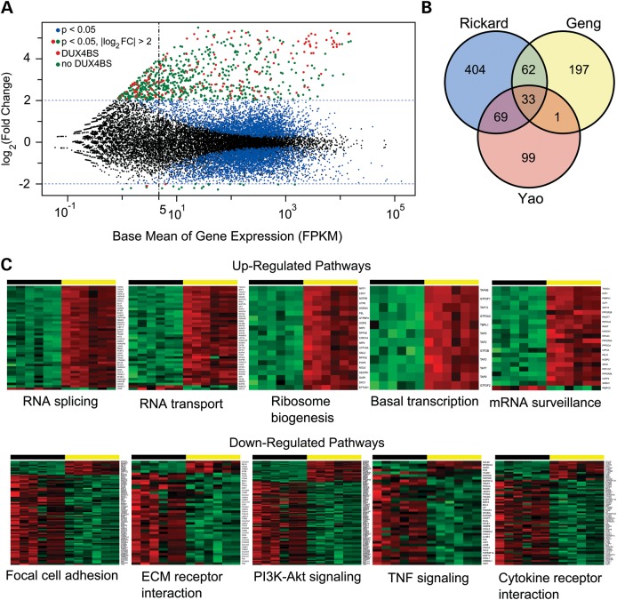
Facioscapulohumeral muscular dystrophy (FSHD) is caused by chromatin relaxation that results in aberrant expression of the transcription factor Double Homeobox 4 (DUX4). DUX4 protein is present in a small subset of FSHD muscle cells, making its detection and analysis of its effects historically difficult. Using a DUX4-activated reporter, we demonstrate the burst expression pattern of endogenous DUX4, its method of signal amplification in the unique shared cytoplasm of the myotube, and FSHD cell death that depends on its activation. Transcriptome analysis of DUX4-expressing cells revealed that DUX4 activation disrupts RNA metabolism including RNA splicing, surveillance and transport pathways. Cell signaling, polarity and migration pathways were also disrupted. Thus, DUX4 expression is sufficient for myocyte death, and these findings suggest mechanistic links between DUX4 expression and cell migration, supporting recent descriptions of phenotypic similarities between FSHD and an FSHD-like condition caused by FAT1 mutations.


Figures

Figure 1.
Figure 1.
Characterization of a DUX4-activated fluorescent reporter. (A) A schematic diagram of the lentiviral vector encoding a DUX4 reporter with five unique DUX4 binding site sequences, identified from individual DUX4 genomic targets and a TATA box located upstream of the sequence for nuclear-localized Blue Fluorescent Protein (nuBFP). A pTK and neomycin phosphotransferase gene (NEO) were included to allow for selection of transduced cells. (B) BFP is specifically present in MHC-positive myotubes (green) derived from individuals with FSHD and co-localizes with DUX4 protein (red) by immunofluorescence. (C) Delivery of an siRNA targeting the DUX4 transcript (siDUX4) during FSHD myoblast differentiation prohibits BFP reporter activation by eliminating DUX4 protein expression. A universal non-targeting control siRNA (siCTRL) transfected in parallel results in BFP and DUX4 expression typical of FSHD myotubes. (D) qRT-PCR of DUX4 and DUX4 targets BFP, CCNA1 and MBD3L2 shows reduced mRNA levels after siDUX4 delivery during FSHD myoblast differentiation. DUX4 and DUX4 target mRNA levels are typical of FSHD cells when siDUX4 is substituted with siCTRL. (N.D., not detected). (E) Immunostaining of endogenous DUX4 target ZNF217 (red) co-localizes with BFP fluorescence (blue) during FSHD myoblast differentiation.
Figure 2.
Figure 2.
DUX4 and target expression reveal temporal stages of the transcriptional cascade resulting from DUX4 activation. (A) Four distinct patterns identified by line scan fluorescence intensity analysis suggest that DUX4-activated target expression follows transient asynchronous pulses of DUX4 expression and can persist even after DUX4 is no longer detectable. (B) DUX4 protein originates from sentinel DUX4+ nuclei that do not express DUX4 targets (1), then diffuses to nearby nuclei in the shared cytoplasm of the myotube and activates target genes (2). This momentary expression of DUX4 followed by diffusion results in a gradient of the protein and its targets (3), followed by sustained target expression in nuclei previously exposed to DUX4 (4).
Figure 3.
Figure 3.
Imaging and quantification of DUX4 target activation and cell death events during FSHD myoblast differentiation. (A) Brightfield and GFP live cell imaging of differentiating FSHD myoblasts associates DUX4-activated reporter expression with cell death (white arrows) occurring ∼20.2 h after GFP is detectable (data not shown). (B) Quantification of DUX4-activated reporter detection in sentinel events and associated cell death events during 120 h of FSHD myoblast differentiation, where each graph describes an independent differentiation experiment and reports total events observed over 11.7 mm2.
Figure 4.
Figure 4.
Quantification of DUX4 activation events and DUX4 and DUX4 target mRNA expression analysis in sorted myoblasts. (A) Representative flow cytometry plots of BFP fluorescence intensity (X-axis) versus autofluorescence (Y-axis) in 72 h differentiated control and FSHD myoblasts harboring the DUX4-activated BFP reporter with % reporter + cells displayed for each line. (B) Populations of BFP+ and BFP– myoblasts were collected by flow sorting and mRNA levels of DUX4, and DUX4 targets BFP, CCNA1 and MBD3L2 were measured by qRT-PCR. (N.D., not detected).
Figure 5.
Figure 5.
RNA-seq analysis of differentially expressed genes in DUX4-expressing cells. (A) Scatter plot of the log2FC in gene expression verses the mean expression level for each gene. All points from three biological replicates of two different FSHD myoblast cell lines are shown (6 samples sorted for DUX4-expressing and DUX4-non-expressing cells, 12 samples total). Gene expression levels with significant (P < 0.05) log2FC values are shown as colored dots (blue, red and green). Data points for genes with log2FC change >2 (linear fold change >4) are colored in red if they contain a DUX4 binding site within 5 kb of the transcription start site and green if they do not. DUX4 binding sites were determined by analysis of publically available ChIP-seq data (13). (B) Venn diagram comparing DUX4 mis-expressed genes found in previous RNA-seq studies (Geng and Yao) to those identified here. (C) Examples of several significantly dysregulated gene sets listed in Table 1. Differentially expressed genes identified using the DESeq2 R package and plotted in (A) were used for Generally Applicable Gene-set/Pathway Analysis (GAGE) (25). The genes from a particular pathway that showed significant up- or down-regulation are displayed as heat maps (green low, red high) for all 12 data points. As in (A), three biological replicates of two FSHD cell lines are shown, and data points for DUX4 reporter-negative cells (black horizontal bar) and DUX4 reporter-positive cells (yellow horizontal bar) are shown as different color shades based on the regularized log transformation (log2(counts)) of normalized counts. Gene symbols are listed vertically along the right border of each heat map.

References

    1. Deenen J.C., Arnts H., van der Maarel S.M., Padberg G.W., Verschuuren J.J., Bakker E., Weinreich S.S., Verbeek A.L., van Engelen B.G. (2014) Population-based incidence and prevalence of facioscapulohumeral dystrophy. Neurology, 83, 1056–1059. - PMC - PubMed
    1. Tawil R. (2008) Facioscapulohumeral muscular dystrophy. Neurotherapeutics, 5, 601–606. - PMC - PubMed
    1. Taylor D.A., Carroll J.E., Smith M.E., Johnson M.O., Johnston G.P., Brooke M.H. (1982) Facioscapulohumeral dystrophy associated with hearing loss and Coats syndrome. Ann. Neurol., 12, 395–398. - PubMed
    1. Gurwin E.B., Fitzsimons R.B., Sehmi K.S., Bird A.C. (1985) Retinal telangiectasis in facioscapulohumeral muscular dystrophy with deafness. Arch. Ophthalmol., 103, 1695–1700. - PubMed
    1. van Overveld P.G., Lemmers R.J., Sandkuijl L.A., Enthoven L., Winokur S.T., Bakels F., Padberg G.W., van Ommen G.J., Frants R.R., van der Maarel S.M. (2003) Hypomethylation of D4Z4 in 4q-linked and non-4q-linked facioscapulohumeral muscular dystrophy. Nat. Genet., 35, 315–317. - PubMed

Publication types

MeSH terms