

E-cadherin-defective gastric cancer cells depend on Laminin to survive and invade
- PMID: 26246502
- PMCID: PMC4581612
- DOI: 10.1093/hmg/ddv312
E-cadherin-defective gastric cancer cells depend on Laminin to survive and invade
Abstract
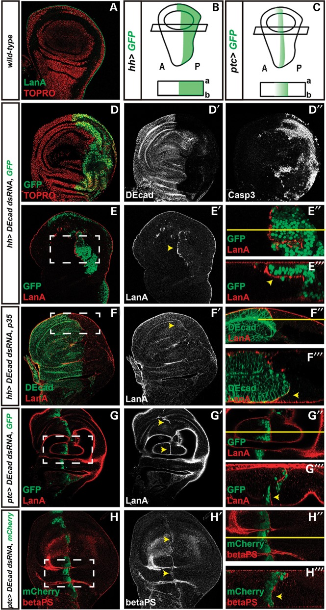
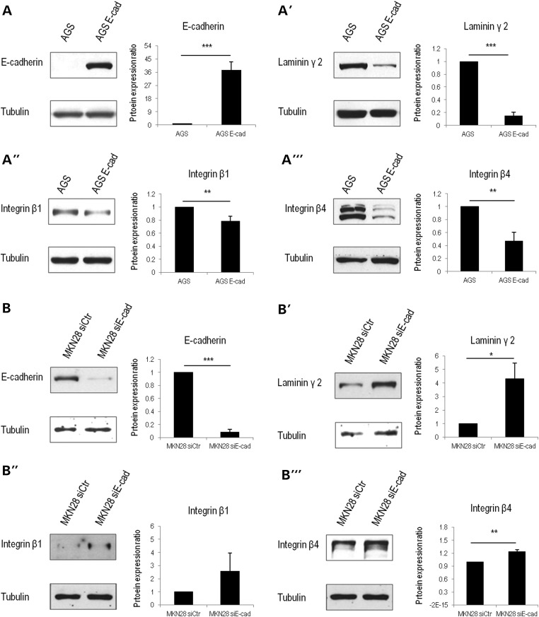
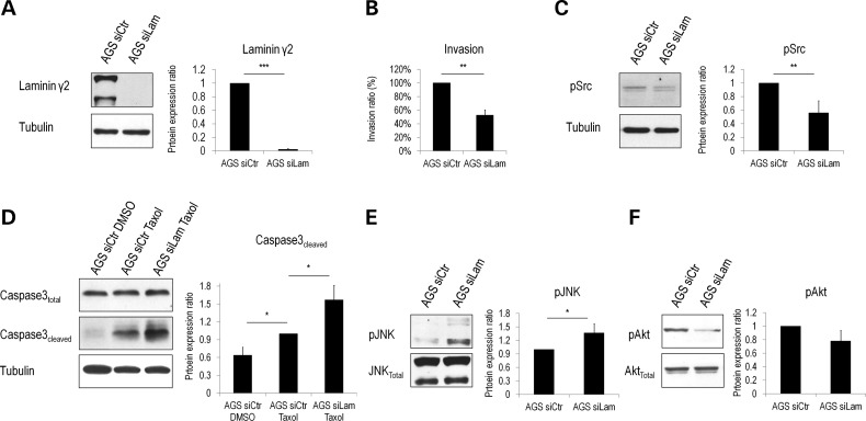
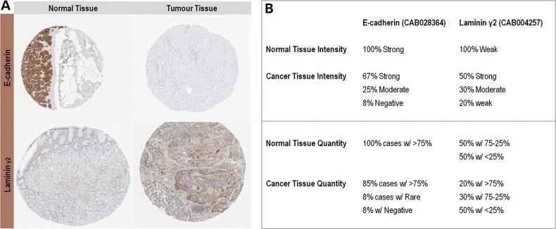
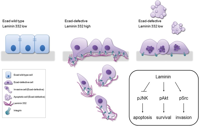
Epithelial-cadherin (Ecad) deregulation affects cell-cell adhesion and results in increased invasiveness of distinct human carcinomas. In gastric cancer, loss of Ecad expression is a common event and is associated with disease aggressiveness and poor prognosis. However, the molecular mechanisms underlying the invasive process associated to Ecad dysfunction are far from understood. We hypothesized that deregulation of cell-matrix interactions could play an important role during this process. Thus, we focussed on LM-332, which is a major matrix component, and in Ecad/LM-332 crosstalk in the process of Ecad-dependent invasion. To verify whether matrix deregulation was triggered by Ecad loss, we used the Drosophila model. To dissect the key molecules involved and unveil their functional significance, we used gastric cancer cell lines. The relevance of this relationship was then confirmed in human primary tumours. In vivo, Ecad knockdown induced apoptosis; nonetheless, at the invasive front, cells ectopically expressed Laminin A and βPS integrin. In vitro, we demonstrated that, in two different gastric cancer cell models, Ecad-defective cells overexpressed Laminin γ2 (LM-γ2), β1 and β4 integrin, when compared with Ecad-competent ones. We showed that LM-γ2 silencing impaired invasion and enhanced cell death, most likely via pSrc and pAkt reduction, and JNK activation. In human gastric carcinomas, we found a concomitant decrease in Ecad and increase in LM-γ2. This is the first evidence that ectopic Laminin expression depends on Ecad loss and allows Ecad-dysfunctional cells to survive and invade. This opens new avenues for using LM-γ2 signalling regulators as molecular targets to impair gastric cancer progression.
© The Author 2015. Published by Oxford University Press. All rights reserved. For Permissions, please email: journals.permissions@oup.com.
Figures
Similar articles
-
DNAJB4 molecular chaperone distinguishes WT from mutant E-cadherin, determining their fate in vitro and in vivo.Hum Mol Genet. 2014 Apr 15;23(8):2094-105. doi: 10.1093/hmg/ddt602. Epub 2013 Nov 29. Hum Mol Genet. 2014. PMID: 24293545
-
Co-expression of laminin β3 and γ2 chains and epigenetic inactivation of laminin α3 chain in gastric cancer.Int J Oncol. 2011 Sep;39(3):593-9. doi: 10.3892/ijo.2011.1048. Epub 2011 May 20. Int J Oncol. 2011. PMID: 21617852
-
Laminin gamma2 mediates Wnt5a-induced invasion of gastric cancer cells.Gastroenterology. 2009 Jul;137(1):242-52, 252.e1-6. doi: 10.1053/j.gastro.2009.02.003. Gastroenterology. 2009. PMID: 19582886
-
Invasion and metastases in gastric cancer: in vitro and in vivo models with clinical correlations.Semin Oncol. 1996 Jun;23(3):316-24. Semin Oncol. 1996. PMID: 8658215 Review.
-
Therapeutic targets associated to E-cadherin dysfunction in gastric cancer.Expert Opin Ther Targets. 2013 Oct;17(10):1187-201. doi: 10.1517/14728222.2013.827174. Epub 2013 Aug 19. Expert Opin Ther Targets. 2013. PMID: 23957294 Review.
Cited by
-
Clinical spectrum and pleiotropic nature of CDH1 germline mutations.J Med Genet. 2019 Apr;56(4):199-208. doi: 10.1136/jmedgenet-2018-105807. Epub 2019 Jan 19. J Med Genet. 2019. PMID: 30661051 Free PMC article. Review.
-
Clinical importance of E-cadherin deficiency in resectable gastric cancer: A nested case-control study.Oncol Lett. 2025 Jul 4;30(3):427. doi: 10.3892/ol.2025.15173. eCollection 2025 Sep. Oncol Lett. 2025. PMID: 40688580 Free PMC article.
-
Multicellular Human Gastric-Cancer Spheroids Mimic the Glycosylation Phenotype of Gastric Carcinomas.Molecules. 2018 Oct 30;23(11):2815. doi: 10.3390/molecules23112815. Molecules. 2018. PMID: 30380716 Free PMC article.
-
The Extracellular Matrix: An Accomplice in Gastric Cancer Development and Progression.Cells. 2020 Feb 8;9(2):394. doi: 10.3390/cells9020394. Cells. 2020. PMID: 32046329 Free PMC article. Review.
-
Differential Impacts on Tensional Homeostasis of Gastric Cancer Cells Due to Distinct Domain Variants of E-Cadherin.Cancers (Basel). 2022 May 29;14(11):2690. doi: 10.3390/cancers14112690. Cancers (Basel). 2022. PMID: 35681670 Free PMC article.
References
-
- Paredes J., Figueiredo J., Albergaria A., Oliveira P., Carvalho J., Ribeiro A.S., Caldeira J., Costa A.M., Simoes-Correia J., Oliveira M.J. et al. (2012) Epithelial E- and P-cadherins: role and clinical significance in cancer. Biochim. Biophys. Acta, 1826, 297–311. - PubMed
-
- Nottingham J. (1994) Signet-ring carcinoma of stomach in a child. Histopathology, 24, 490–491. - PubMed
-
- Corso G., Carvalho J., Marrelli D., Vindigni C., Carvalho B., Seruca R., Roviello F., Oliveira C. (2013) Somatic mutations and deletions of the E-cadherin gene predict poor survival of patients with gastric cancer. J. Clin. Oncol., 31, 868–875. - PubMed
-
- Takeichi M. (1993) Cadherins in cancer: implications for invasion and metastasis. Curr. Opin. Cell Biol., 5, 806–811. - PubMed
Publication types
MeSH terms
Substances
Grants and funding
LinkOut - more resources
Full Text Sources
Other Literature Sources
Medical
Molecular Biology Databases
Research Materials
