. 2015 Nov;89(21):11129-43.
doi: 10.1128/JVI.01612-15. Epub 2015 Aug 5.

A Druggable Pocket at the Nucleocapsid/Phosphoprotein Interaction Site of Human Respiratory Syncytial Virus

Affiliations

A Druggable Pocket at the Nucleocapsid/Phosphoprotein Interaction Site of Human Respiratory Syncytial Virus

Mohamed Ouizougun-Oubari et al. J Virol. 2015 Nov.

Abstract

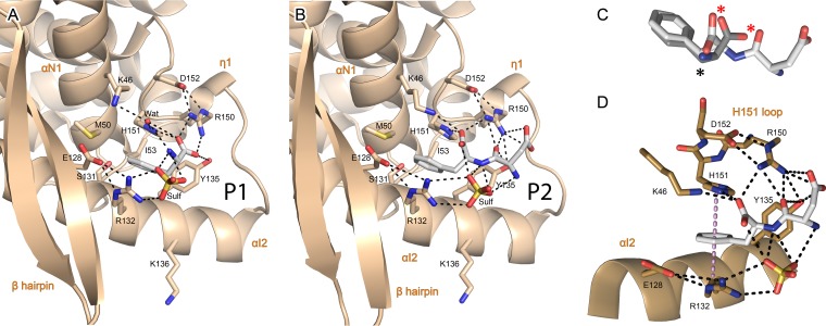
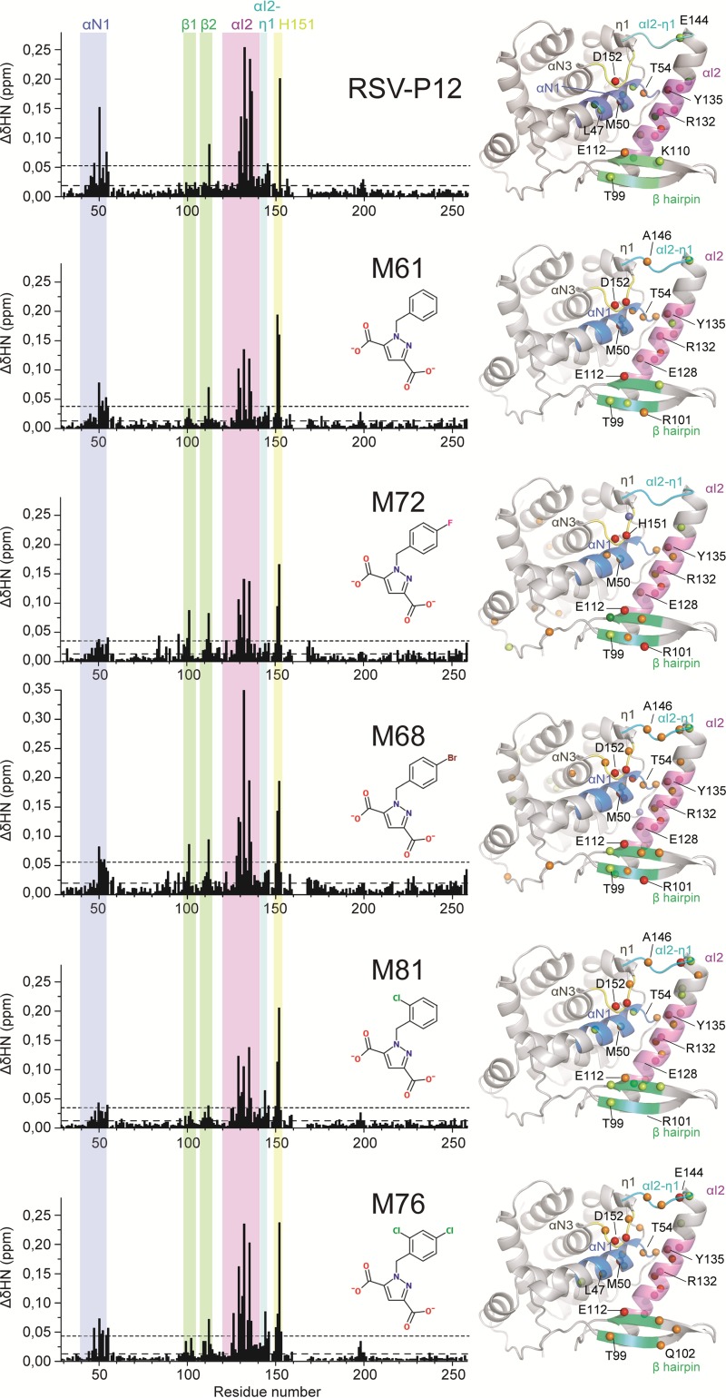
Presently, respiratory syncytial virus (RSV), the main cause of severe respiratory infections in infants, cannot be treated efficiently with antivirals. However, its RNA-dependent polymerase complex offers potential targets for RSV-specific drugs. This includes the recognition of its template, the ribonucleoprotein complex (RNP), consisting of genomic RNA encapsidated by the RSV nucleoprotein, N. This recognition proceeds via interaction between the phosphoprotein P, which is the main polymerase cofactor, and N. The determinant role of the C terminus of P, and more particularly of the last residue, F241, in RNP binding and viral RNA synthesis has been assessed previously. Here, we provide detailed structural insight into this crucial interaction for RSV polymerase activity. We solved the crystallographic structures of complexes between the N-terminal domain of N (N-NTD) and C-terminal peptides of P and characterized binding by biophysical approaches. Our results provide a rationale for the pivotal role of F241, which inserts into a well-defined N-NTD pocket. This primary binding site is completed by transient contacts with upstream P residues outside the pocket. Based on the structural information of the N-NTD:P complex, we identified inhibitors of this interaction, selected by in silico screening of small compounds, that efficiently bind to N and compete with P in vitro. One of the compounds displayed inhibitory activity on RSV replication, thereby strengthening the relevance of N-NTD for structure-based design of RSV-specific antivirals.

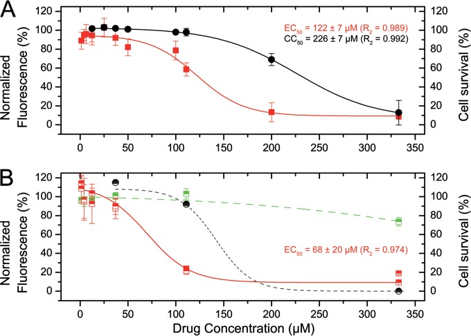
Importance: Respiratory syncytial virus (RSV) is a widespread pathogen that is a leading cause of acute lower respiratory infections in infants worldwide. RSV cannot be treated efficiently with antivirals, and no vaccine is presently available, with the development of pediatric vaccines being particularly challenging. Therefore, there is a need for new therapeutic strategies that specifically target RSV. The interaction between the RSV phosphoprotein P and the ribonucleoprotein complex is critical for viral replication. In this study, we identified the main structural determinants of this interaction, and we used them to screen potential inhibitors in silico. We found a family of molecules that were efficient competitors of P in vitro and showed inhibitory activity on RSV replication in cellular assays. These compounds provide a basis for a pharmacophore model that must be improved but that holds promises for the design of new RSV-specific antivirals.


Figures

FIG 1
FIG 1
X-ray structures of N-NTD and P-CTD binding sites. (A) Superposition of isolated N-NTDs in a free form (pale and bright orange, chains A and B for both crystal forms) or in complex with a P C-terminal peptide, P2 (light gray), and N-NTD in the RSV RNP context (black; PDB 2WJ8) in cartoon representation. Secondary structures are labeled according to reference . The P C-terminal peptide P2 is shown in stick representation colored by atom type, with carbons in white. (B to E) Final 2Fo-Fc electron density map for P1 (B), P2 (C), M76 (D), and M72 (E) at 2.0-, 2.4-, 2.1-, and 2.9-Å X-ray resolutions, respectively. The maps are drawn in blue mesh contoured at 1.0 sigma, with the ligand displayed as sticks.
FIG 2
FIG 2
Details of the P-CTD binding pocket and interaction network with the N-NTD. (A and B) Enlarged views of the binding of phenylalanine (P1) (A) and Asp-Phe (P2) (B), corresponding to the P C terminus. The N-NTD is in tan and shown in cartoon representation, with secondary structures labeled. Residues involved in binding through electrostatic (dashed lines) or van der Waals interactions are shown in stick representation and colored by atom type, with carbons in tan. P1 and P2 are in stick representation, with the same color scheme as in Fig. 1. (C) Superposition of P2 and P1 conformations. Atoms with conserved polar contacts are indicated by asterisks, notably, two oxygen atoms (red asterisks). (D) Details of the double stacking interaction between the aromatic ring of P2 and the side chains of H151 and R132, with electrostatic interactions between P2 and the N-NTD indicated by black dashed lines. The normal axis to the P-F24 ring plane is indicated by the violet dotted line.
FIG 3
FIG 3
NMR interaction experiments using 1H-15N HSQC spectra of the N-NTD. (A) 1H-15N HSQC spectrum at 800 MHz 1H frequency of 50 μM 15N-labeled N-terminal domain of RSV nucleoprotein at 293 K. Peak assignments are indicated according to the numbering in the native sequence. Unassigned amide or side chain peaks are indicated by asterisks. The inset (top left) shows detailed assignment of the central region of the spectrum (boxed area). Asn and Gln NH2 side chain resonances are connected by horizontal lines. (B) Enlarged views of superimposed 1H-15N HSQC spectra obtained by titrating either RSV-P12 peptide (top) or M76 (bottom) into 15N N-NTD. Chemical shift perturbations of large amplitude are indicated by arrows. Molar ligand/protein ratios and color codes are indicated on the right of each spectrum.
FIG 4
FIG 4
(Left) Mapping of chemical shift perturbations induced by P peptide and BPdC ligands. CSP profiles were extracted from 1H-15N HSQC spectra of 15N-labeled N-NTD, with the P12 peptide and BPdC ligands at the titration midpoint, where CSPs were half of those at saturation. The bar graphs represent combined CSPs containing both 1H and 15N contributions (Δδ1H15N). Mean values (long dashes) and means plus 1 standard deviation (short dashes) are plotted for each ligand. (Right) CSPs were mapped onto the 3D structure of the N-NTD. Amide nitrogen atoms of residues with large CSPs are represented as spheres, with a color code reflecting the contributions of 1H and 15N chemical shifts: gray, Δδ1H > 0.06 ppm; dark green, Δδ15N > 0.3 ppm; light green, Δδ15N > 0.2 ppm; red; Δδ1H15N > 0.09 ppm; and orange, Δδ1H15N > 0.06 ppm. The regions where most CSPs are observed are labeled on the diagrams and on the structure: the C terminus of helix αN1 in blue, the center of helix αI2 in magenta, the αI2-η1 loop in cyan, the H151-loop in yellow, and the β-hairpin in green.
FIG 5
FIG 5
X-ray structures of N-NTD:BPdC complexes. (A and B) Enlarged view of the binding of M76 (A) and M72 (B) in the N-NTD pocket. The N-NTD is displayed as in Fig. 1, with M76 and M72 in stick representation colored by atom type, with magenta and yellow carbons, respectively. Stabilizing electrostatic interactions are displayed as black dashed lines. The insets show superposition of M76 and P2, and a black star marks the most deeply buried atom of both ligands in the binding pocket (A) and superposition of M76 and M72 interacting with S131 through a halogen bond (yellow dashed line) and an H bond (black dashed line) (B). (A, C, and D) The green arrow indicates the water molecule in head-on interaction with the para Cl. (C) Details of the double stacking interaction involving the H151 imidazole, the benzene ring of M76, and the R132 side chain. H bonds and halogen bonds are plotted as in panels A and B. The normal axis to the benzene ring plane of M76 running through H151 NE2 and R132 NE is shown as a violet dotted line. (D) Electrostatic interactions of the two halogen substituents in M76, with H bonds and halogen bonds displayed as in panels A and B. The final 2Fo-Fc (blue) and Fo-Fc (red) electron density maps are displayed around M76 at 2σ and 3σ, respectively. Additional intraprotein H bonds stabilizing N-NTD secondary-structure elements and involved in M76 binding or involving direct contacts between solvent molecules and M76 are displayed in light blue.
FIG 6
FIG 6
N-NTD surface complementarity for P peptide and BPdC ligands. Shown are enlarged views of the binding of P2 (A), M76 (B), and M72 (C). The N-NTD van der Waals surface is represented according to the electrostatic potential, with positive (blue) and negative (red) potentials shown. Ligands are displayed as van der Waals spheres and colored by atom type, with white carbons.
FIG 7
FIG 7
BPdC competition with P-CTD for N-NTD binding by SPR. (Top) Real-time association and dissociation profiles corresponding to the injection over immobilized P-CTD of the N-NTD premixed with different concentrations of M61 (left) or M76 (right). (Bottom) BPdC concentration dependence of the steady-state SPR response upon N-NTD injection over a P-CTD surface relative to the response in the absence of BPdC. The lines represent the best fit of the experimental data to a single class of binding site model.
FIG 8
FIG 8
Inhibition of rHRSV-mCherry replication by M76-diAM. (A) HEp-2 cells in 96-well plates were infected with 500 PFU of rHRSV-mCherry in the presence of serial dilutions of M76-diAM. The red fluorescence (red symbols) and the luminescence reporting on cell survival (black symbols) were read at 48 h postinfection by automatic counting and normalized based on the fluorescence and luminescence of nontreated infected cells. Each data point represents the mean of the results of three experiments done in triplicate. The standard errors of the mean (SEM) are represented by the error bars. The lines correspond to the data fitted to a dose-response curve. (B) BHK-21 cells in 96-well plates were infected with 500 PFU of rHRSV-mCherry or 50 PFU VSV-GFP in the presence of serial dilutions of M76-diAM. Red (red symbols) and green (green symbols) fluorescence, as well as cell survival (black symbols), were read at 48 h postinfection by automatic counting and normalized based on the fluorescence and luminescence of nontreated infected cells. Each data point represents the mean (±SEM) of two experiments done in triplicate. rHRSV-mCherry fluorescence was fitted to a dose-response curve (solid line). The approximate fit of VSV-GFP fluorescence and cytotoxicity to dose response curves are plotted with green and black dashed lines, respectively.
FIG 9
FIG 9
Evolutionary conservation of the N-NTD binding pocket in the RNP context. (Top) Surface representation of N-NTD colored according to the evolutionary conservation of amino acids, calculated using the ConSurf server (45), with turquoise-to-magenta indicating variable-to-conserved. The result is displayed as the crystallographic model of the RSV RNP (PDB 2WJ8 [16]) restricted to the N-NTD, without terminal arms or the N-CTD. The two neighboring N-NTD protomers in the RNP are displayed in cartoon (tan). The RNA molecule and M76 are displayed in stick form and colored by atom type, with white and magenta carbons, respectively. (Bottom) Location of the P-CTD binding site on the model of the authentic left-handed helical RNP (PDB 4BKK [3]). The N-NTD (tan), N-CTD (yellow), and terminal arms (gray) are shown. The RNA molecule and M76 are shown as white sticks and ball and stick colored as in panel A, respectively. In the N-NTD, residues corresponding to N mutants with a minigenome replication activity of <33%, <66%, or >133% that of the wild type are colored red, orange, and green, respectively (15). On the right is an enlarged view of the N-NTD.

Similar articles

Cited by

References

    1. Nair H, Nokes DJ, Gessner BD, Dherani M, Madhi SA, Singleton RJ, O'Brien KL, Roca A, Wright PF, Bruce N, Chandran A, Theodoratou E, Sutanto A, Sedyaningsih ER, Ngama M, Munywoki PK, Kartasasmita C, Simoes EA, Rudan I, Weber MW, Campbell H. 2010. Global burden of acute lower respiratory infections due to respiratory syncytial virus in young children: a systematic review and meta-analysis. Lancet 375:1545–1555. doi:10.1016/S0140-6736(10)60206-1. - DOI - PMC - PubMed
    1. Olszewska W, Openshaw P. 2009. Emerging drugs for respiratory syncytial virus infection. Expert Opin Emerg Drugs 14:207–217. doi:10.1517/14728210902946399. - DOI - PMC - PubMed
    1. Bakker SE, Duquerroy S, Galloux M, Loney C, Conner E, Eleouet JF, Rey FA, Bhella D. 2013. The respiratory syncytial virus nucleoprotein-RNA complex forms a left-handed helical nucleocapsid. J Gen Virol 94:1734–1738. doi:10.1099/vir.0.053025-0. - DOI - PMC - PubMed
    1. Collins PL, Hill MG, Camargo E, Grosfeld H, Chanock RM, Murphy BR. 1995. Production of infectious human respiratory syncytial virus from cloned cDNA confirms an essential role for the transcription elongation factor from the 5′ proximal open reading frame of the M2 mRNA in gene expression and provides a capability for vaccine development. Proc Natl Acad Sci U S A 92:11563–11567. doi:10.1073/pnas.92.25.11563. - DOI - PMC - PubMed
    1. Yu Q, Hardy RW, Wertz GW. 1995. Functional cDNA clones of the human respiratory syncytial (RS) virus N, P, and L proteins support replication of RS virus genomic RNA analogs and define minimal trans-acting requirements for RNA replication. J Virol 69:2412–2419. - PMC - PubMed

Publication types