. 2015 Oct;89(20):10333-46.
doi: 10.1128/JVI.00991-15. Epub 2015 Aug 5.

Hepatitis C Virus Envelope Glycoprotein E1 Forms Trimers at the Surface of the Virion

Affiliations

Hepatitis C Virus Envelope Glycoprotein E1 Forms Trimers at the Surface of the Virion

Pierre Falson et al. J Virol. 2015 Oct.

Abstract

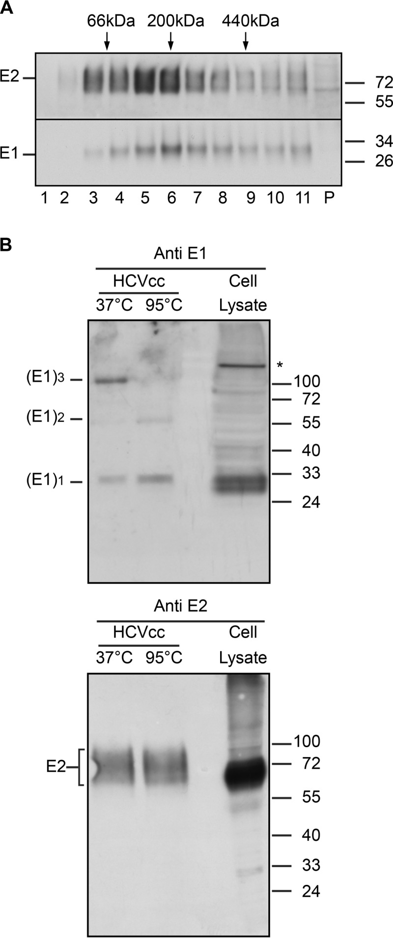
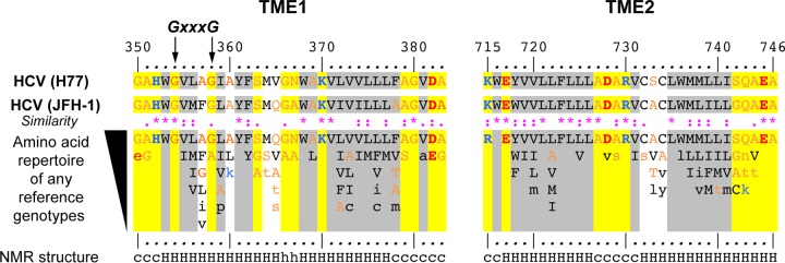
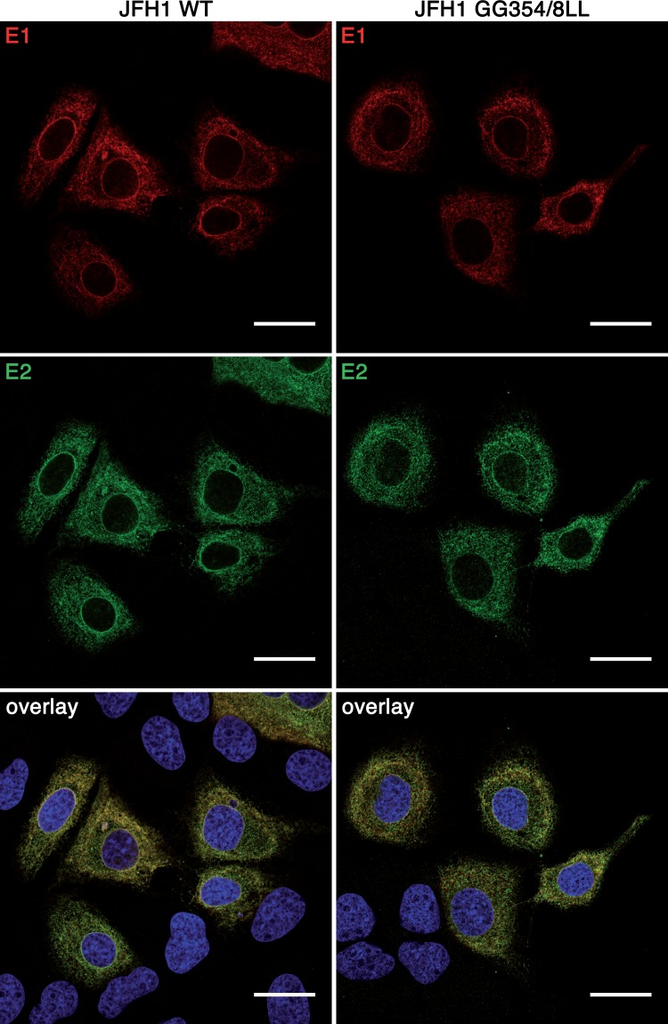
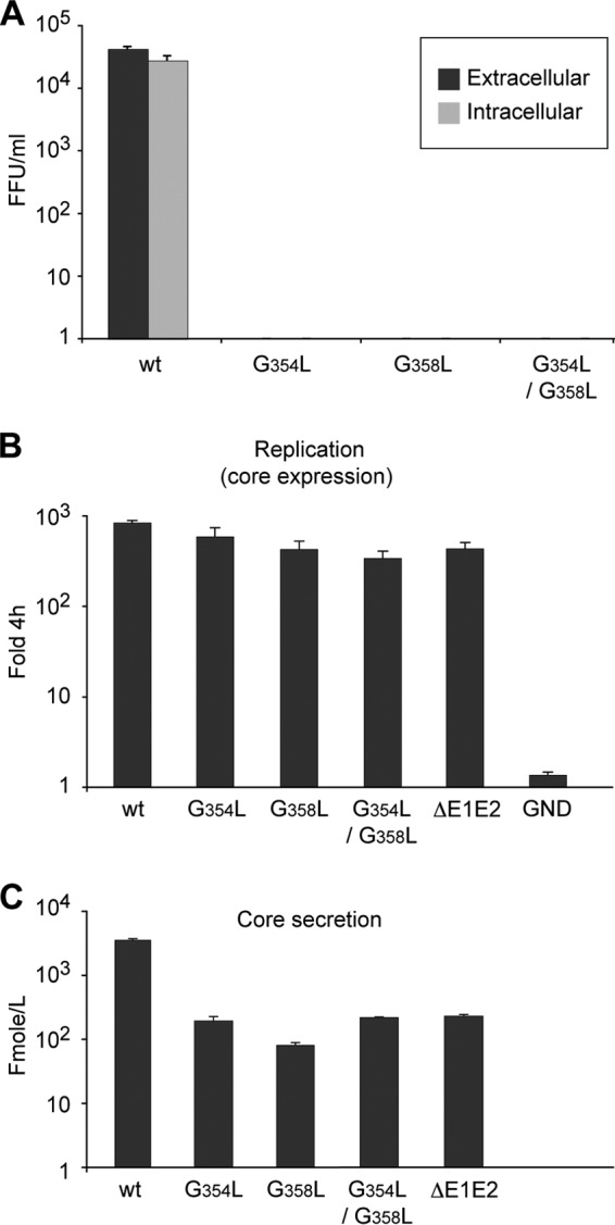
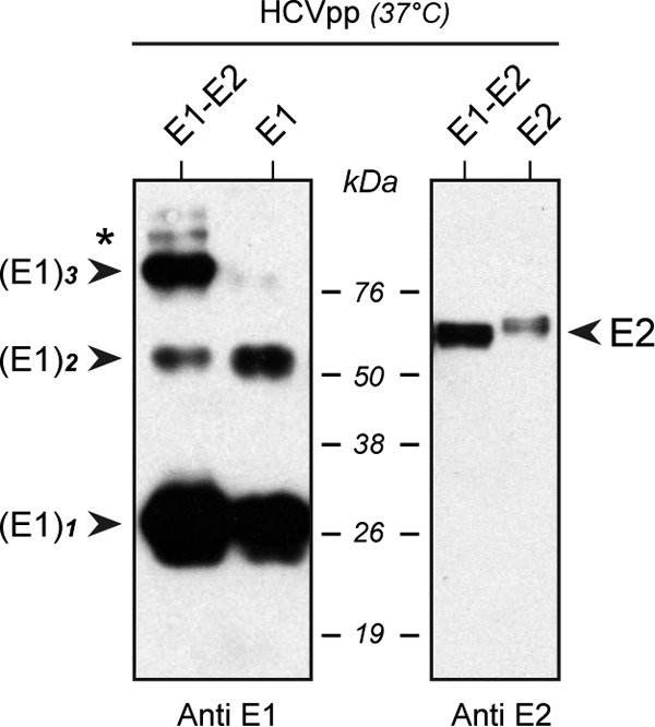
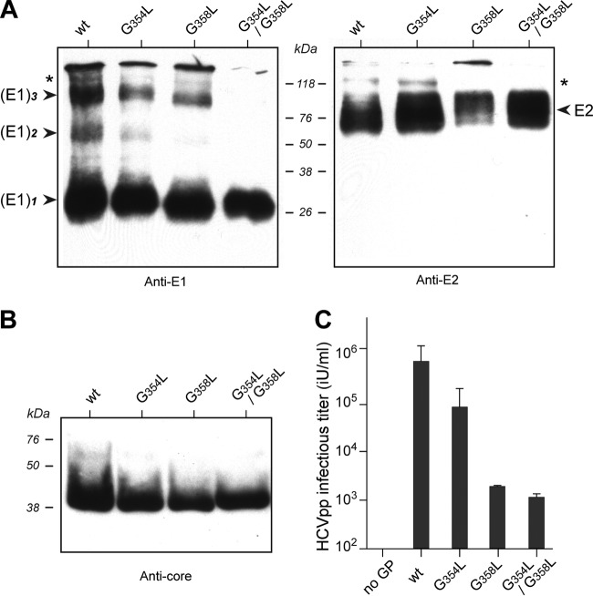
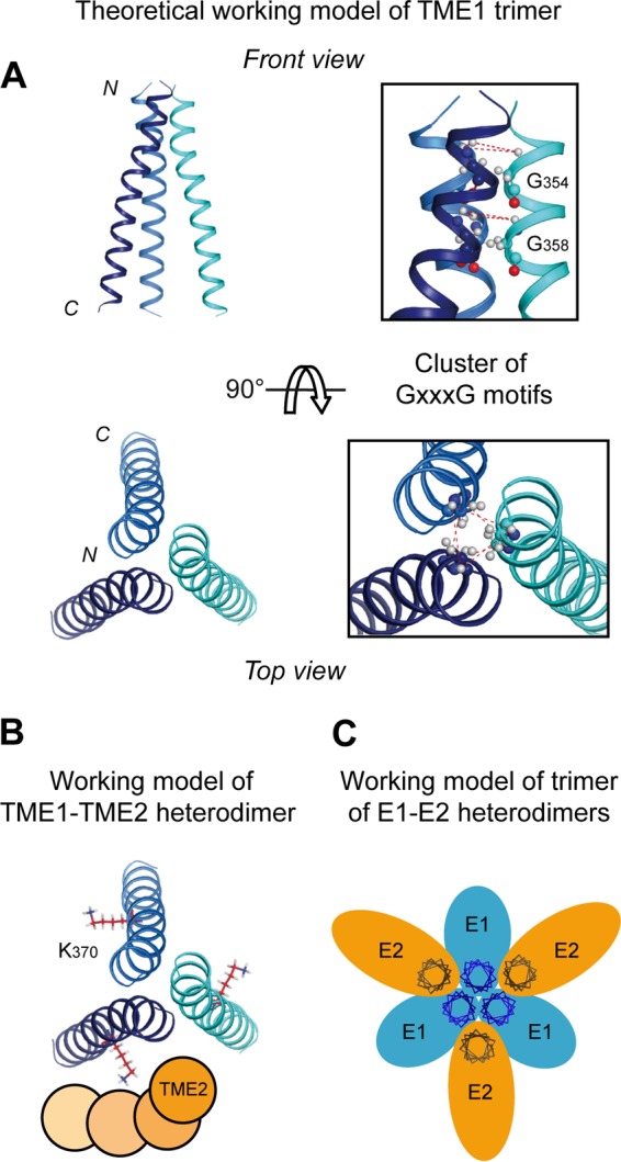
In hepatitis C virus (HCV)-infected cells, the envelope glycoproteins E1 and E2 assemble as a heterodimer. To investigate potential changes in the oligomerization of virion-associated envelope proteins, we performed SDS-PAGE under reducing conditions but without thermal denaturation. This revealed the presence of SDS-resistant trimers of E1 in the context of cell-cultured HCV (HCVcc) as well as in the context of HCV pseudoparticles (HCVpp). The formation of E1 trimers was found to depend on the coexpression of E2. To further understand the origin of E1 trimer formation, we coexpressed in bacteria the transmembrane (TM) domains of E1 (TME1) and E2 (TME2) fused to reporter proteins and analyzed the fusion proteins by SDS-PAGE and Western blotting. As expected for strongly interacting TM domains, TME1-TME2 heterodimers resistant to SDS were observed. These analyses also revealed homodimers and homotrimers of TME1, indicating that such complexes are stable species. The N-terminal segment of TME1 exhibits a highly conserved GxxxG sequence, a motif that is well documented to be involved in intramembrane protein-protein interactions. Single or double mutations of the glycine residues (Gly354 and Gly358) in this motif markedly decreased or abrogated the formation of TME1 homotrimers in bacteria, as well as homotrimers of E1 in both HCVpp and HCVcc systems. A concomitant loss of infectivity was observed, indicating that the trimeric form of E1 is essential for virus infectivity. Taken together, these results indicate that E1E2 heterodimers form trimers on HCV particles, and they support the hypothesis that E1 could be a fusion protein.

Importance: HCV glycoproteins E1 and E2 play an essential role in virus entry into liver cells as well as in virion morphogenesis. In infected cells, these two proteins form a complex in which E2 interacts with cellular receptors, whereas the function of E1 remains poorly understood. However, recent structural data suggest that E1 could be the protein responsible for the process of fusion between viral and cellular membranes. Here we investigated the oligomeric state of HCV envelope glycoproteins. We demonstrate that E1 forms functional trimers after virion assembly and that in addition to the requirement for E2, a determinant for this oligomerization is present in a conserved GxxxG motif located within the E1 transmembrane domain. Taken together, these results indicate that a rearrangement of E1E2 heterodimer complexes likely occurs during the assembly of HCV particles to yield a trimeric form of the E1E2 heterodimer. Gaining structural information on this trimer will be helpful for the design of an anti-HCV vaccine.


Figures

FIG 1
FIG 1
Analysis of HCV envelope glycoproteins associated with viral particles. (A) Separation of HCV envelope glycoproteins in a sucrose density gradient. HCV glycoproteins from a semipurified viral preparation were lysed in 1% Triton X-100 and were separated by sedimentation through a 5-to-20% sucrose gradient in the presence of DTT. Eleven 1-ml fractions and the gradient pellet (P) were harvested. After GNA pulldown, samples were analyzed by reducing SDS-PAGE and Western blotting for the presence of E1 and E2 glycoproteins. The sedimentation profiles of several standard globular proteins in a parallel gradient are indicated above the gel: BSA (66 kDa), β-amylase (200 kDa), and ferritin (440 kDa). (B) Analyses of HCV envelope glycoproteins by SDS-PAGE without heat denaturation. HCVcc particles were lysed in 1% Triton X-100, and HCV envelope glycoproteins were pulled down with a GST-CD81 fusion protein. The proteins were treated with Laemmli sample buffer and were heated for 5 min at 37°C (no thermal denaturation) or 95°C (thermal denaturation) before separation by SDS-PAGE. HCV envelope glycoproteins were revealed by Western blotting with anti-E1 MAb 1C4 (top) and anti-E2 MAb 3/11 (bottom). Lysates of HCV-infected cells treated at 95°C were run in parallel. Molecular mass markers (in kilodaltons) are indicated on the right. The oligomeric forms of E1 are indicated on the left. The asterisk indicates the presence of a nonspecific band.
FIG 2
FIG 2
Sequence analyses and NMR structures of the transmembrane domains of HCV E1 and E2. Amino acids of the transmembrane domains of E1 (TME1) (amino acids 350 to 383) and E2 (TME2) (amino acids 715 to 746) are numbered with respect to the HCV polyprotein of the H77 infectious clone (GenBank accession number AF009606) (top). Below the H77 sequence are the sequence of clone JFH-1 (GenBank accession number AB047639) and the amino acid repertoire of the 27 representative TME1 and TME2 sequences from confirmed HCV genotypes and subtypes (listed with accession numbers in Table 1 of reference ; see the European HCV database for details [https://euhcvdb.ibcp.fr/euHCVdb/] [75]). The degree of amino acid conservation at each position can be inferred from the extent of variability (with the observed amino acids listed in decreasing order of frequency from top to bottom) together with the similarity index according to the CLUSTAL W convention (asterisk, invariant; colon, highly similar; dot, similar) (76). Amino acids observed only once at a given position among the 27 sequences are indicated by lowercase letters. To highlight the variable sequence positions in TME1 and TME2, conserved hydrophilic and hydrophobic positions are highlighted in yellow and gray, respectively. Residues are color-coded according to their hydrophobicity: hydrophobic residues are shown in black (Pro, Cys, Val, Leu, Ile, Met, Phe, Tyr, Trp), polar residues in orange (Gly, Ala, Ser, Thr, Asn, Gln), and positively and negatively charged groups of basic (His, Lys, Arg) and acidic (Glu, Asp) residues in blue and red, respectively. The NMR secondary structure (bottom) shows the conformation of residues determined by nuclear magnetic resonance of the N-terminal part of TME1 in 50% trifluoroethanol (7) (PDB entry 1EMZ) and of recombinant TME1 (9) and TME2 (8) in LPPG [1-palmitoyl-2-hydroxy-sn-glycero-3-phospho-(1′-rac-glycerol)] micelles. Residue conformations are indicated as undetermined (c) or helical (H or h; a lowercase h indicates flexible residues [8]). The glycine residues of the GxxxG motif that were mutated individually (G354L or G358L) or together (G354L G358L) to leucine in the TM domain of E1 are indicated by arrows.
FIG 3
FIG 3
Effect of the mutation of the GxxxG motif on E1 trimerization. Glycine residues of the GxxxG motif in the TM domain of E1 were replaced individually (G354L or G358L) or together (G354L/G358L) by leucine in the context of the HCVcc system. Huh-7 cells were electroporated with HCV RNA transcribed in vitro, incubated for 48 h at 37°C, and lysed in 1% Triton X-100. (A) HCV envelope glycoproteins were pulled down with GNA, treated with Laemmli sample buffer, and heated for 5 min at 37°C before separation by SDS-PAGE. HCV envelope glycoproteins were revealed by Western blotting with anti-E1 MAb A4 and anti-E2 MAb 3/11. The oligomeric forms of E1 are indicated on the left and the putative E1E2 heterodimer on the right. The asterisk on the right shows an additional band revealed with the anti-E2 antibody, which likely corresponds to the uncleaved E2p7NS2 precursor. (B) In a parallel experiment, HCV envelope glycoproteins were pulled down with the GST-CD81 fusion protein, treated with Laemmli sample buffer, and heated for 5 min at 70°C before separation by SDS-PAGE. The ratio of coprecipitated E1 glycoprotein to E2 was measured by quantifying the bands by densitometry.
FIG 4
FIG 4
Mutation of the GxxxG motif in E1 does not affect its subcellular localization. Huh-7 cells were electroporated with HCV RNA transcribed in vitro. At 48 h posttransfection, cells were fixed and were processed for immunofluorescence with MAbs A4 (anti-E1) and AR3A (anti-E2). Bars, 25 μm.
FIG 5
FIG 5
Effects of mutation of the GxxxG motif on HCV infectivity and assembly. In vitro-transcribed RNAs of the GxxxG mutants were electroporated into Huh-7 cells. At 72 h postelectroporation, intracellular and extracellular infectivities (A), expressed in focus-forming units (FFU) per milliliter, were determined by titration, and the amounts of intracellular (B) and extracellular (C) core antigen were measured. Note that the amount of intracellular core protein is expressed as the fold increase in core expression over the level measured at 4 h postelectroporation. The ΔE1E2 mutant, which includes an in-frame deletion in the E1E2 region, and the GND mutant, which includes a mutation in the active site of the viral polymerase, were used as negative controls for virus assembly and replication, respectively.
FIG 6
FIG 6
Analysis of HCV envelope glycoproteins associated with HCV pseudoparticles. HCVpp displaying wild-type E1 and E2 complexes, or only E1 or E2, were pelleted through 20% sucrose cushions and were resuspended in PBS. The concentrated viral suspension was treated with Laemmli sample buffer and was heated for 5 min at 37°C prior to separation by SDS-PAGE. HCV envelope glycoproteins were revealed by Western blotting with monoclonal antibodies A4 and H52, directed against E1 and E2, respectively. (Left) The formation of E1 trimers is dependent on the presence of E2 in HCVpp. (Right) The absence of E1 on HCVpp strongly reduces the expression of E2. Homo- and hetero-oligomeric species of E1 and E2 are indicated. The asterisk on the left indicates an additional band that might correspond to the E1E2 heterodimer.
FIG 7
FIG 7
The GxxxG motif in TME1 is required for E1 trimerization and HCVpp infectivity. (A) HCVpp displaying wild-type E2 in combination with wild-type E1 (wt) or with E1 displaying the G354L or G358L mutation, or the G354L G358L double mutation, were concentrated on sucrose cushions and resuspended in PBS. The corresponding HCVpp suspensions were treated with Laemmli sample buffer and were heated for 5 min at 37°C prior to analysis by SDS-PAGE and immunoblotting with anti-E1 antibody A4 or anti-E2 antibody H52. The asterisk indicates an additional band that might correspond to the E1E2 heterodimer. (C) The infectivity of HCV pseudoparticles was measured by titrating the unconcentrated viral supernatants onto Huh-7 cells. Virions contained a viral GFP marker gene that leads to the expression of GFP in successfully infected target cells. Titers are expressed as GFP transducing units per milliliter. (B) The amount of viral capsid in each viral supernatant was also quantified by immunoblotting with an anti-capsid antibody.
FIG 8
FIG 8
The TM domain of E1 (TME1) expressed in E. coli forms GxxxG-dependent trimers and TME1–TME2 oligomers. (Left) Bacteria expressing thioredoxin (Trx) fused to wild-type TME1 (Trx-TME1) or to TME1 displaying either the G354L, G358L, or G354L and G358L (G354/358L) mutations were lysed, heated for 5 min at 37°C in Laemmli sample buffer, and subjected to SDS-PAGE, followed by immunoblotting with an anti-Trx antibody. Monomeric, dimeric, and trimeric forms of TME1 are indicated. (Right) Coexpression of Trx-TME1 and GST-TME2 (GST fused to TME2) in E. coli. Bacteria expressing a discistronic construct encoding Trx-TME1 and GST-TME2 were induced and were subjected to SDS-PAGE analysis and immunoblotting with an anti-Trx antibody. Homo- and hetero-oligomeric forms of Trx-TME1 and GST-TME2 are indicated.
FIG 9
FIG 9
Theoretical models of the TME1 trimer and the trimer of E1E2 heterodimers. (A) Ribbon representation of the trimer model of TME1. Note that, for clarity, TME2 is not represented (see the legend to panel B below). This symmetric model was constructed by assuming the implication of glycine 354 and glycine 358 of each monomer at the trimer interfaces. Dashed red lines indicate interhelical amide proton distances used in the modeling protocol. Glycine residues 354 and 358 forming the GxxxG motifs are shown in ball-and-stick representations. (B) Theoretical model of TME1–TME2 interaction assuming a Lys370 (TME1)–Asp728 (TME2) salt bridge and an Asn367 (TME1)–Asp728 (TME2) interhelical hydrogen bond (as proposed in reference 37). Because numerous possible model solutions were obtained for the relative positioning of TME2 to TME1, TME2 is schematically represented as orange circles at four representative positions for a single TME1–TME2 heterodimer. (C) Schematic representation of a possible organization of the trimer of E1E2 heterodimers at the membrane surface of the viral particle. E1 and E2 ectodomains are represented as blue and yellow ovals, respectively, while TME1 and TME2 are represented as helix projections (i.e., perpendicular to the membrane surface). These models were generated from structure coordinates using VMD (http://www.ks.uiuc.edu/Research/vmd/) and were rendered with POV-Ray (http://www.povray.org/).

Similar articles

Cited by

References

    1. Stapleton JT, Foung S, Muerhoff AS, Bukh J, Simmonds P. 2011. The GB viruses: a review and proposed classification of GBV-A, GBV-C (HGV), and GBV-D in genus Pegivirus within the family Flaviviridae. J Gen Virol 92:233–246. doi:10.1099/vir.0.027490-0. - DOI - PMC - PubMed
    1. Adams MJ, King AM, Carstens EB. 2013. Ratification vote on taxonomic proposals to the International Committee on Taxonomy of Viruses (2013). Arch Virol 158:2023–2030. doi:10.1007/s00705-013-1688-5. - DOI - PubMed
    1. Moradpour D, Penin F, Rice CM. 2007. Replication of hepatitis C virus. Nat Rev Microbiol 5:453–463. doi:10.1038/nrmicro1645. - DOI - PubMed
    1. Vieyres G, Dubuisson J, Pietschmann T. 2014. Incorporation of hepatitis C virus E1 and E2 glycoproteins: the keystones on a peculiar virion. Viruses 6:1149–1187. doi:10.3390/v6031149. - DOI - PMC - PubMed
    1. Cocquerel L, Wychowski C, Minner F, Penin F, Dubuisson J. 2000. Charged residues in the transmembrane domains of hepatitis C virus glycoproteins play a key role in the processing, subcellular localization, and assembly of these envelope proteins. J Virol 74:3623–3633. doi:10.1128/JVI.74.8.3623-3633.2000. - DOI - PMC - PubMed

Publication types

MeSH terms

Substances