. 2015 Oct;89(20):10489-99.
doi: 10.1128/JVI.00304-15. Epub 2015 Aug 5.

Evasion of the Innate Immune Type I Interferon System by Monkeypox Virus

Affiliations

Evasion of the Innate Immune Type I Interferon System by Monkeypox Virus

William D Arndt et al. J Virol. 2015 Oct.

Abstract

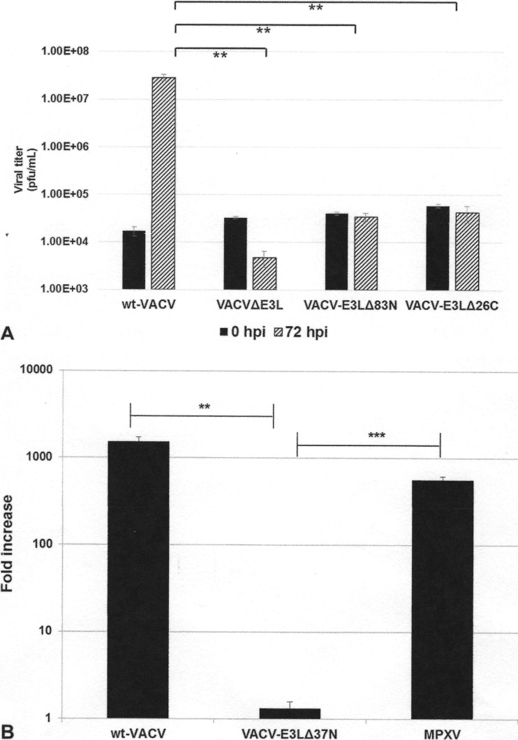
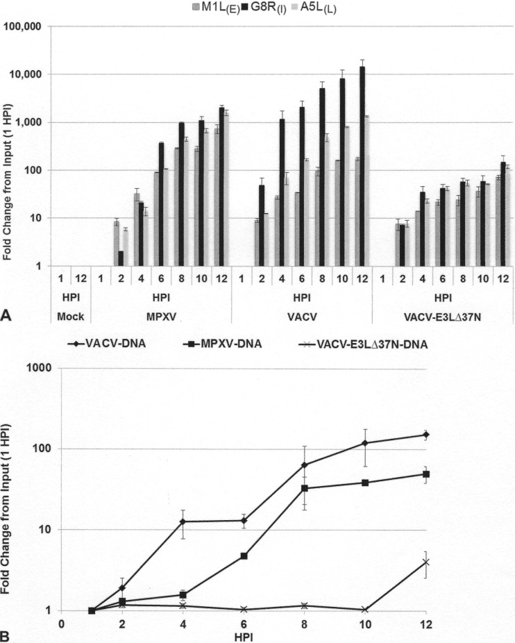
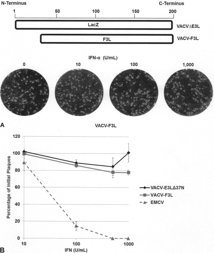
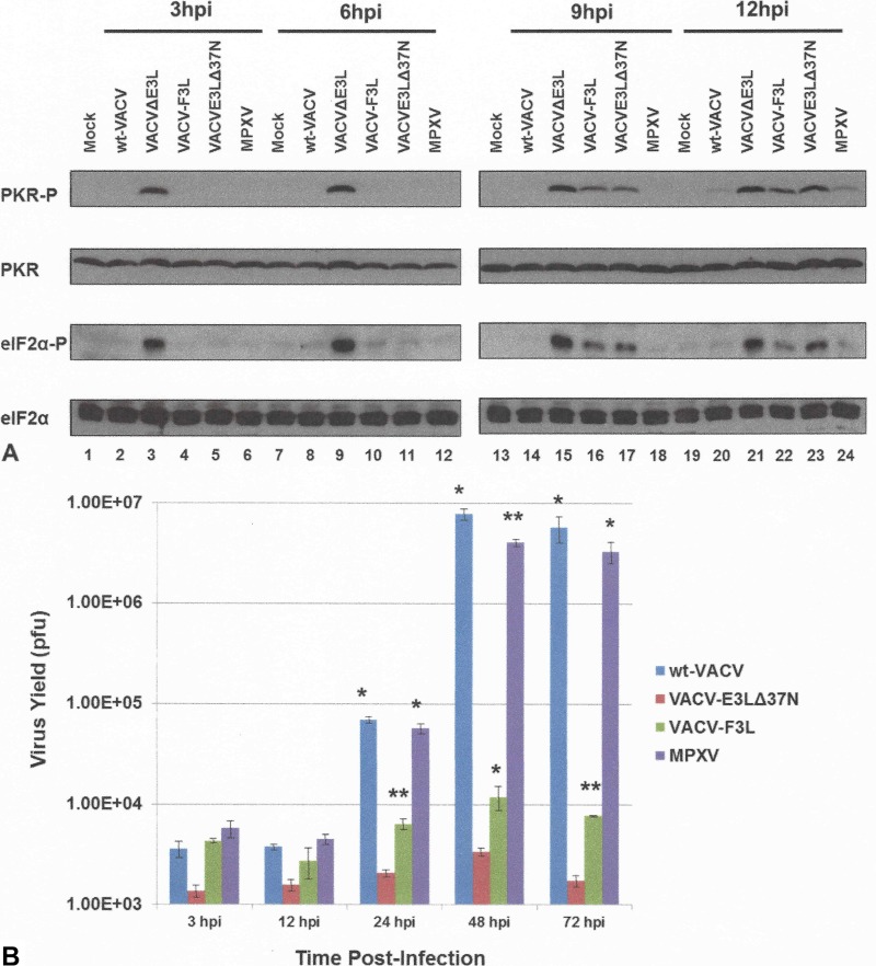
The vaccinia virus (VACV) E3 protein has been shown to be important for blocking activation of the cellular innate immune system and allowing viral replication to occur unhindered. Mutation or deletion of E3L severely affects viral host range and pathogenesis. While the monkeypox virus (MPXV) genome encodes a homologue of the VACV E3 protein, encoded by the F3L gene, the MPXV gene is predicted to encode a protein with a truncation of 37 N-terminal amino acids. VACV with a genome encoding a similarly truncated E3L protein (VACV-E3LΔ37N) has been shown to be attenuated in mouse models, and infection with VACV-E3LΔ37N has been shown to lead to activation of the host antiviral protein kinase R pathway. In this report, we present data demonstrating that, despite containing a truncated E3 homologue, MPXV phenotypically resembles a wild-type (wt) VACV rather than VACV-E3LΔ37N. Thus, MPXV appears to contain a gene or genes that can suppress the phenotypes associated with an N-terminal truncation in E3. The suppression maps to sequences outside F3L, suggesting that the suppression is extragenic in nature. Thus, MPXV appears to have evolved mechanisms to minimize the effects of partial inactivation of its E3 homologue.

Importance: Poxviruses have evolved to have many mechanisms to evade host antiviral innate immunity; these mechanisms may allow these viruses to cause disease. Within the family of poxviruses, variola virus (which causes smallpox) is the most pathogenic, while monkeypox virus is intermediate in pathogenicity between vaccinia virus and variola virus. Understanding the mechanisms of monkeypox virus innate immune evasion will help us to understand the evolution of poxvirus innate immune evasion capabilities, providing a better understanding of how poxviruses cause disease.


Figures

FIG 1
FIG 1
Schematic of viruses used in this study.
FIG 2
FIG 2
(A) VACV-E3L homologue alignment. The first 160 nucleotides of the VACV E3L gene homologues of VACV, VARV, and MPXV were aligned by use of the ClustalW program. The sequences begin at nucleotide 1 of each gene, which is the start codon in the case of VACV and VARV. The C-terminal dsRNA BD is highly conserved among all three viruses. Nonhomologous nucleotide differences in MPXV that lead to the inability to generate p25 are shown in gray boxes. The conserved AUG codon that leads to the generation of p20 (the only product expressed by MPXV) is boxed only. Asterisks indicate the nucleotide positions of the consensus sequence among all three sequences. (B) Protein sequence alignment. The VACV E3 protein homologues of VACV, VARV, and MPXV were aligned by use of the ClustalW program. Asterisks, amino acid positions of the consensus sequence among all three sequences; amino acids highlighted in gray, changes in sequences among MPXV, VARV, and VACV; dots, degree of similarity, with one dot being less conservative than two. The N-terminal Z-NA BD for VACV and VARV is highly conserved, whereas the E3 homologue of MPXV (F3) is predicted to contain a 37-amino-acid N-terminal truncation. (C) MPXV produces a p20 form of VACV-E3. HeLa cells were infected with wt VACV, VACV-E3LΔ37N, VACV-F3L, and MPXV at an MOI of 5. At 3 (lanes 2 to 6), 6 (lanes 7 to 11), 9 (lanes 13 to 17), and 12 (lanes 18 to 22) hpi, protein lysates were isolated and analyzed by Western blotting with antibodies specific for the C terminus of E3.
FIG 3
FIG 3
MPXV replication in the presence of IFN. (A) RK13 cells were treated with increasing amounts of IFN-α A/D for 18 h and then infected with 100 PFU of MPXV. Cells were stained at 48 hpi with crystal violet. (B) Subconfluent BSC-40 cells were treated with the indicated amount of recombinant IFN for 18 h prior to infection. Treated cells were infected with approximately 100 PFU of VACV, EMCV, or MPXV. Cells were stained with crystal violet at 48 hpi.
FIG 4
FIG 4
Inhibition of PKR pathway by MPXV. HeLa cells were either mock infected (lanes 1 and 2) or infected with wt VACV (lanes 3 to 6), VACVΔE3L (lanes 7 to 10), VACV-E3LΔ37N (lanes 11 to 14), or MPXV (lanes 15 to 18) in the presence (+) (lanes 2, 5, 6, 9, 10, 13, 14, 17, and 18) or absence (lanes 1, 3, 4, 7, 8, 11, 12, 15, and 16) of 1,000 U/ml of IFN at an MOI of 5. Protein lysates were isolated at 6 and 9 hpi and analyzed by Western blotting with antibodies specific to the phosphorylated forms of PKR (PKR-P) and eIF2α (eIF2α-P). Detection of GAPDH was used to ensure equal loading of proteins.
FIG 5
FIG 5
Replication in JC cells. (A) JC cells were infected with the indicated viruses at an MOI of 0.01 PFU/cell. Infected cells were harvested at 0 and 72 hpi, and the titer was determined by plaque assay in RK-E3L cells. Data are presented as means with standard errors from three experiments. **, P ≤ 0.01. (B) JC cells were either mock infected or infected with wt VACV, VACV E3LΔ37N, or MPXV at an MOI of 0.01 PFU/cell. Viruses were harvested at 72 hpi, and the titer was determined by plaque assay in BSC-40 cells. Data represent the fold increase in viral growth at 72 hpi and are presented as means with standard errors from multiple experiments. Statistical analyses were done by using an unpaired t test comparing wt VACV and MPXV to VACV-E3LΔ37N. **, P ≤ 0.01; ***, P ≤ 0.001.
FIG 6
FIG 6
(A) Gene expression in JC cells. JC cells were either mock infected or infected with MPXV, VACV-E3LΔ37N, or VACV at an MOI of 5. RNA was extracted at 1, 2, 4, 6, 8, 10, and 12 hpi. Real-time quantitative PCR was performed with primers specific for M1L (early [E]), G8R (intermediate [I), and A5L (late [L]) viral gene transcripts. The transcript level of each gene was graphed as the fold change from the amount input (at 1 hpi). Data represent means with standard errors from multiple experiments. (B) Genomic replication in JC cells. Cells were infected with VACV, VACV-E3LΔ37N, or MPXV at an MOI of 5. Cells were harvested at 1, 2, 4, 6, 8, 10, and 12 hpi, and total DNA was extracted with phenol chloroform. Total DNA (500 ng) was used for real-time PCR with VACV G8R gene-specific primers. The graph presents the fold change in genomic DNA as the fold change from the amount input (at 1 hpi), along with transcript levels from Fig. 5A. Data represent means with standard errors from multiple experiments.
FIG 7
FIG 7
The MPXV F3 protein restores the IFNr phenotype. (A) Through in vitro recombination, the F3L gene of MPXV was inserted into the E3L locus of VACVΔE3L, generating a recombinant VACV that expresses the MPXV F3 protein (VACV-F3L). RK13 cells were treated with increasing amounts of IFN-α A/D for 18 h, prior to infection with 100 PFU of VACV-F3L. At 48 hpi, the cells were stained with crystal violet. (B) Subconfluent BSC-40 cells were treated with the indicated amount of recombinant IFN at 18 hpi. Treated cells were infected with approximately 100 PFU of VACV E3LΔ37N, VACV-F3L, or EMCV. Cells were stained with crystal violet at 48 hpi.
FIG 8
FIG 8
(A) Late activation of the PKR pathway by VACV-F3L. HeLa cells were either mock infected (lanes 1, 7, 13, and 19) or infected with wt VACV (lanes 2, 8, 14, and 20), VACVΔE3L (lanes 3, 9, 15, and 21), VACV-F3L (lanes 4, 10, 16, and 22), VACV-E3LΔ37N (lanes 5, 11, 17, and 23), or MPXV (lanes 6, 12, 18, and 24) at an MOI of 5. Protein lysates were isolated at 3, 6, 9, and 12 hpi and analyzed by Western blotting with antibodies specific to the phosphorylated forms of PKR and eIF2α. (B) VACV-F3L replication is inhibited in JC cells. JC cells were infected with wt VACV, VACV E3LΔ37N, VACV-F3L, or MPXV at an MOI of 0.01 PFU/cell. Viruses were harvested at 3, 12, 24, 48, and 72 hpi, and the titer in BSC-40 cells was determined by plaque assay. Data represent means with standard errors from multiple experiments. Statistical significance was determined by comparison of the results for each group against those for VACV-E3LΔ37N using a multiple t test. *, P ≤ 0.05; **, P ≤ 0.01.

References

    1. Marennikova SS, Seluhina EM, Mal'ceva NN, Cimiskjan KL, Macevic GR. 1972. Isolation and properties of the causal agent of a new variola-like disease (monkeypox) in man. Bull World Health Organ 46:599–611. - PMC - PubMed
    1. Chen N, Li G, Liszewski MK, Atkinson JP, Jahrling PB, Feng Z, Schriewer J, Buck C, Wang C, Lefkowitz EJ, Esposito JJ, Harms T, Damon IK, Roper RL, Upton C, Buller RM. 2005. Virulence differences between monkeypox virus isolates from West Africa and the Congo Basin. Virology 340:46–63. doi:10.1016/j.virol.2005.05.030. - DOI - PMC - PubMed
    1. Huhn GD, Bauer AM, Yorita K, Graham MB, Sejvar J, Likos A, Damon IK, Reynolds MG, Kuehnert MJ. 2005. Clinical characteristics of human monkeypox, and risk factors for severe disease. Clin Infect Dis 41:1742–1751. doi:10.1086/498115. - DOI - PubMed
    1. Fenner F, Henderson DA, Arita I, Jezek Z, Ladnyi ID. 1988. Smallpox and its eradication, vol 6 World Health Organization, Geneva, Switzerland.
    1. Reed KD, Melski JW, Graham MB, Regnery RL, Sotir MJ, Wegner MV, Kazmierczak JJ, Stratman EJ, Li Y, Fairley JA, Swain GR, Olson VA, Sargent EK, Kehl SC, Frace MA, Kline R, Foldy SL, Davis JP, Damon IK. 2004. The detection of monkeypox in humans in the Western Hemisphere. N Engl J Med 350:342–350. doi:10.1056/NEJMoa032299. - DOI - PubMed

Publication types

MeSH terms

LinkOut - more resources