. 2015 Oct 1;26(19):3439-50.
doi: 10.1091/mbc.E15-05-0318. Epub 2015 Aug 5.

Replication stress in early S phase generates apparent micronuclei and chromosome rearrangement in fission yeast

Affiliations

Replication stress in early S phase generates apparent micronuclei and chromosome rearrangement in fission yeast

Sarah A Sabatinos et al. Mol Biol Cell. .

Abstract

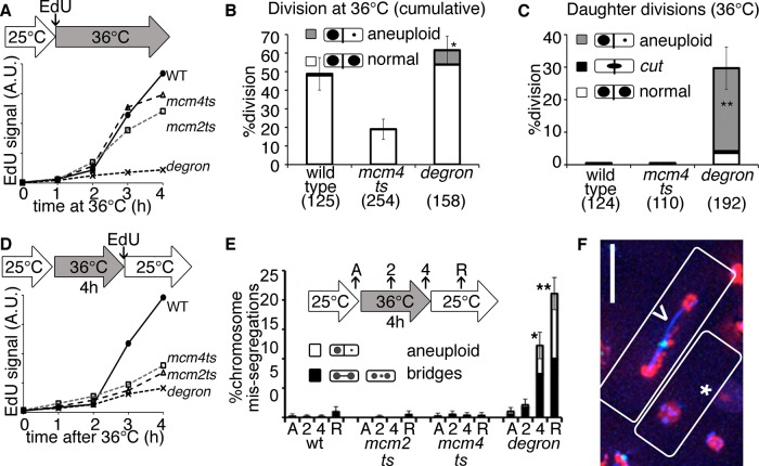
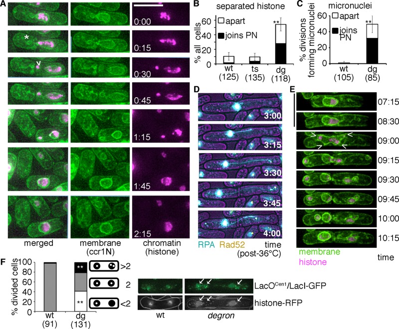
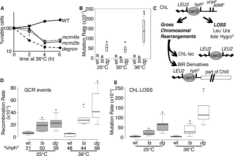
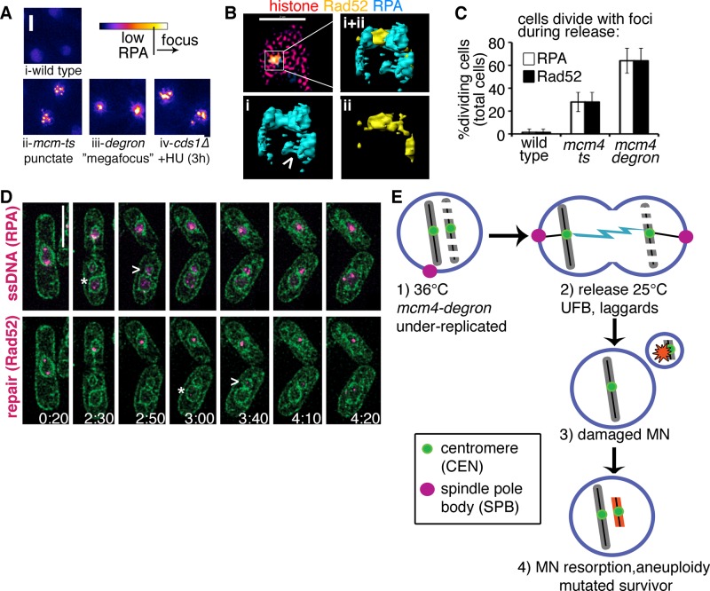
DNA replication stress causes genome mutations, rearrangements, and chromosome missegregation, which are implicated in cancer. We analyze a fission yeast mutant that is unable to complete S phase due to a defective subunit of the MCM helicase. Despite underreplicated and damaged DNA, these cells evade the G2 damage checkpoint to form ultrafine bridges, fragmented centromeres, and uneven chromosome segregations that resembles micronuclei. These micronuclei retain DNA damage markers and frequently rejoin with the parent nucleus. Surviving cells show an increased rate of mutation and chromosome rearrangement. This first report of micronucleus-like segregation in a yeast replication mutant establishes underreplication as an important factor contributing to checkpoint escape, abnormal chromosome segregation, and chromosome instability.


Figures

FIGURE 1:
FIGURE 1:
Underreplicated mcm4-degron mutants divide during and after replication stress. (A) EdU incorporation is lowest in mcm4-degron cells during incubation at 36°C. Asynchronous cultures were shifted to 36°C during EdU exposure, and synthesis was measured by EdU-FACS fluorescence in arbitrary units (A.U.). (B) Wild-type (wt) and mcm4-degron cells divide at least once at 36°C (6-h total videomicroscopy of asynchronous cultures at 36°C). Significantly more mcm4-degron cells undergo reductional anaphase (gray) in this first division at 36°C. Proportions in B, C, and E are shown with 95% CI, Z test of significance (*p < 0.01, **p << 0.001). (C) mcm4-degron daughter cells divide their DNA unevenly at 36°C, whereas wild-type daughters delay division at 36°C. Samples as in B, daughter cell divisions only. (D) Mutants incorporate less EdU after release at 25°C, as measured by EdU FACS. The initial asynchronous population was treated for 4 h at 36°C before release at 25°C in the presence of EdU. (E) Proportion of abnormal nuclear divisions in single-time-point images acquired before heat (asynchronous [A]), at 36°C (2, 4 h), and after 2 h of recovery at 25°C (R). (F) mcm4-degron cells during 25°C release with RPA-CFP (blue), Rad52-YFP (green), and histone-RFP (red). Unequal histone division in one cell (*) and a bridge (>) with a lagging chromosome and repair focus are common. Scale bar, 10 μm.
FIGURE 2:
FIGURE 2:
Underreplication followed by division promotes micronuclei and genomic rearrangement in fission yeast. (A) mcm4-degron cells released to 25°C (after 4 h, 36°C) form anucleate cells (*) and apparent micronuclei (>). Histone-RFP and membrane (ccr1N-GFP) are shown; scale, 10 μm. Micronuclei are often resorbed back into the main nucleus (time 1:15–1:45 h:min) after release to 25°C. (B) Chromatin (histone-RFP) frequently separates into discrete, condensed fragments that are separate from the main nuclear mass in mcm4-degron cells. More than 50% of these fragments rejoin the parent nucleus (PN). Asynchronous cultures were treated (4 h, 36°C) and then shifted to 25°C for microscopy during recovery (12-h cumulative data, two or three biological replicates per strain). The proportion of cells that form separate histone bodies is shown relative to total number of cells monitored. (C) Membrane-enclosed chromatin masses frequently separate from the main nucleus in mcm4-degron cells but are rarely detected in wild type (wt). More than 60% of these micronuclei fuse and rejoin the parent nucleus (PN) during recovery at 25°C (12-h cumulative data, two to four biological replicates per strain). (D) mcm4-degron cells develop dynamic ssDNA (RPA-CFP, blue) bridges dotted with Rad52-YFP (yellow) during release at 25°C (time in hours:minutes). (E) Evidence for two spindles resulting in apparent micronuclei in some mcm4-degron cells during recovery (after 4 h, 36°C). Conditions as in A; scale, 10 μm. (F) A LacO array near the centromere 1 (lys1+-lacOCen1) unevenly separates in mcm4-degron divisions after 4 h at 36°C. More than two dots are frequently observed in mcm4-degron, which suggests that the array is rearranged or fragmented. LacI-GFP (green) bound to lacOCen1 is shown below relative to DNA signal (histone-RFP). Stacked histogram for pooled data from three biological replicates, with chi-squared test of significance for proportion of single dots (gray) or more than three dots (black) segregating (**p << 0.001).
FIGURE 3:
FIGURE 3:
Transient replication instability causes mutation in surviving mcm4-degron cells. (A) Relative viability of cultures at 36°C (strains FY4743, FY4857, FY5279). Cells were shifted to 36°C and plated at time points to determine viability relative to the starting culture. (B) Mutation rate (can1+) increases in mcm4-degron after 4 h at 36°C (n = 7). *p < 0.001 comparing wt or mcm4-ts with mcm4-degron; °p << 0.001 change from 25 to 36°C in mcm4-degron. Plots in B, D, and E show a center median line bounded by 25th and 75th percentiles. (C) Schematic for the minichromosome (ChL) assay, followed by markers (also see Nakamura et al., 2008; Li et al., 2013). Cells may lose ChL or undergo gross chromosomal rearrangements (GCRs). An isochromosome (ChL-iso) is formed by duplication of the left arm with the LEU2+ marker producing a smaller chromosome. Break-induced replication (BIR) products may occur between ChL and chromosome III, producing a longer product that is frequently hygromycin resistant. (D) GCR events are highest in mcm4-degron after 4 h at 36°C. Significant median differences from wild type at 25 or 36°C are reported as p < 0.001 (*) with outliers (o). (E) ChL loss is highest in mcm4-degron after 4 h at 36°C and even before replication stress at 25°C. Loss is also higher in mcm4-degron compared with mcm4-ts (p < 0.02. all conditions). Conditions and analysis as in D.
FIGURE 4:
FIGURE 4:
Divisions occur in the presence of DNA damage and repair signals. (A) RPA focus patterns during replication collapse are different in each mutant but rarely develop in wild type at 36°C (i). Multiple (more than three) punctate RPA foci form in mcm-ts nuclei after 4h 36°C (ii) and later become a pannuclear RPA signal like that observed in cds1∆+HU (iv). A unique “megafocus” of bright, compact RPA forms in mcm4-degron (iii). Heat map scale (top) and 2- μm scale. (B) 3D-SIM images of mcm4-degron nucleus after 4 h 36°C (top left) in one midfocal z-section (xy); scale bar, 2 μm. (i, ii) Enlarged yz-perspectives of surface-rendered megafocus. Also see Supplemental Video S5. (C) RPA and Rad52 foci are present at division in mcm4-degron (also see Supplemental Videos S2 and S6). (D) DNA damage (RPA-CFP, top magenta) and DNA repair foci (Rad52-YFP, bottom magenta) develop in newly formed micronuclei (MN; assessed with membrane marker ccr1N-GFP; green). Cells were incubated at 36°C, 4 h before videomicroscopy during release at 25°C for 6 h. A time scale is indicated on the bottom right corner of each panel (hours:minutes). The MN form damage and repair signals after they are first detected in the parent nucleus (*, parent; >, MN), before rejoining. Scale bar, 5 μm. (E) A proposed model for transient replication-stress inducing mutations in surviving cells after mcm4-degron inactivation, shown at the level of the nucleus (nuclear membrane in blue). Cells treated 4 h at 36°C are underreplicated (step 1) but divide, causing UFBs and fragmented DNA during mitosis (step 2). Fragments are membrane-bound MN that develop DNA damage (step 3). Resorption of MN back into the parent nucleus promotes further genome instability during 25°C recovery, leading to the development of a mutated surviving population (step 4).
FIGURE 5:
FIGURE 5:
Underreplication promotes micronuclei, DNA damage, and aneuploidy in fission yeast. (A) Experimental scheme. Asynchronous cells were shifted to 36°C for 4 h total and then released to 25°C for 2 h. (B) The DNA damage checkpoint becomes activated by Chk1-HA phosphorylation (*) in methyl methanesulfonate (MMS)-treated wild-type cells (+M) and in mcm4-ts. Chk1-HA is moderately phosphorylated in asynchronous mcm4-degron and never attains activated levels of mcm4-ts, as assessed by the ratio of modified (top) to unmodified (bottom) Chk1. The 53BP1 homologue Crb2 is phosphorylated in response to MMS treatment and stable in wild type but is rapidly lost in mcm4-degron at 36°C. Arrowheads (<) indicate modified forms of proteins, and the bar (–) indicates a non–HA-tagged control lysate. (C) Cdc2 is not phosphorylated in mcm4-degron at 36°C and only minimally during recovery (25°C). In contrast, high-level, sustained Cdc2 phosphorylation occurs in mcm4-ts. Cdc2 modified and unmodified protein levels were detected on Western blots and quantified to plot the ratio at each time point. (D) Loss of Mus81 endonuclease (mus81∆) increases divisions in mcm4-ts mus81∆, forming aneuploid and cut cells.

References

    1. Bagley BN, Keane TM, Maklakova VI, Marshall JG, Lester RA, Cancel MM, Paulsen AR, Bendzick LE, Been RA, Kogan SC, et al. A dominantly acting murine allele of mcm4 causes chromosomal abnormalities and promotes tumorigenesis. PLoS Genet. 2012;8:e1003034. - PMC - PubMed
    1. Bailis JM, Luche DD, Hunter T, Forsburg SL. Minichromosome maintenance proteins interact with checkpoint and recombination proteins to promote s-phase genome stability. Mol Cell Biol. 2008;28:1724–1738. - PMC - PubMed
    1. Bartkova J, Horejsi Z, Koed K, Kramer A, Tort F, Zieger K, Guldberg P, Sehested M, Nesland JM, Lukas C, et al. DNA damage response as a candidate anti-cancer barrier in early human tumorigenesis. Nature. 2005;434:864–870. - PubMed
    1. Bass KL, Murray JM, O’Connell MJ. Brc1-dependent recovery from replication stress. J Cell Sci. 2012;125:2753–2764. - PMC - PubMed
    1. Beeharry N, Rattner JB, Caviston JP, Yen T. Centromere fragmentation is a common mitotic defect of S and G2 checkpoint override. Cell Cycle. 2013;12:1588–1597. - PMC - PubMed

Publication types

MeSH terms

Substances

LinkOut - more resources