

DACH1 Expresison in Osteosarcoma and Its Relationship with Proliferation and Angiogenesis
- PMID: 26246702
- PMCID: PMC4522252
- DOI: 10.1007/s12262-012-0761-8
DACH1 Expresison in Osteosarcoma and Its Relationship with Proliferation and Angiogenesis
Abstract
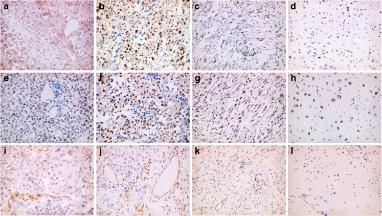
The aim of this study was to investigate the expression of DACH1 in osteosarcoma as well as its relationship with cell proliferation and angiogenesis in the tumor. DACH1 expression was detected by immunohistochemical staining in the serial sections of the osteosarcoma. The microvessel density (MVD) was counted by CD34 immunohistochemical staining, and immunohistochemical staining of PCNA staining showed the cell proliferation. The impacts of DACH1 expression on tumor proliferation and angiogenesis were evaluated by statistics. The DACH1 had different expression patterns in different osteosarcoma. Conventional osteosarcoma showed stronger DACH1 staining (conventional vs. parosteal: P = 0.037; conventional vs. periosteal: P = 0.028) and more PCNA-positive tumor cells than parosteal and periosteal osteosarcoma (conventional vs. parosteal: P = 0.041; conventional vs. periosteal: P = 0.045), the difference was significant. In addition, conventional osteosarcoma showed more cytoplasmic staining of DACH1 than parosteal and periosteal (conventional vs. parosteal: P = 0.023; conventional vs. periosteal: P = 0.030). Parosteal and periosteal osteosarcoma showed no significant difference in DACH1 expression and cell proliferation index. On the other hand, DACH1 different expression patterns showed significantly different impacts on angiogenesis. In spite of the different subtypes of osteosarcoma, the MVD showed a significant difference in cytoplasmic and nuclear expression patterns of DACH1 (nuclear expression vs. cytoplasmic expression: 5.72 ± 1.19 vs. 9.65 ± 1.24, P = 0.042). Moreover, in the conventional osteosarcoma, the MVD also showed a significant difference in DACH1 cytoplasmic and nuclear staining (nuclear expression vs. cytoplasmic expression: 5.58 ± 0.71 vs. 13.65 ± 1.30, P = 0.019). However, the DACH1 expression intensity showed no significant different impacts on MVD of all kinds of osteosarcoma. DACH1 had different expression patterns and intensity. Cytoplasmic and nuclear expression of DACH1 might play different roles in cell proliferation and angiogenesis of osteosarcoma. Cytoplasmic DACH1 might promote cell proliferation and be associated with angiogenesis.
Keywords: Angiogenesis; Cell proliferation; DACH1; Osteosarcoma.
Figures
References
-
- Mardon G, Solomon NM, Rubin GM. Dachshund encodes a nuclear protein required for normal eye and leg development in Drosophila. Development. 1994;120:3473–3486. - PubMed
-
- Shen W, Mardon G. Ectopic eye development in drosophila induced by directed dachsund expression. Development. 1997;124:45–52. - PubMed
LinkOut - more resources
Full Text Sources
Miscellaneous