Review
. 2015:2015:421746.
doi: 10.1155/2015/421746. Epub 2015 Jul 13.

Biology of Bone Tissue: Structure, Function, and Factors That Influence Bone Cells

Affiliations
Review

Biology of Bone Tissue: Structure, Function, and Factors That Influence Bone Cells

Rinaldo Florencio-Silva et al. Biomed Res Int. 2015.

Abstract

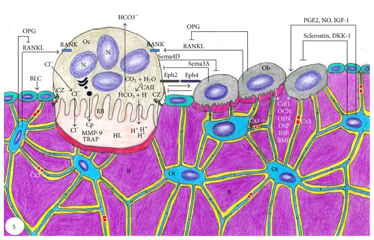
Bone tissue is continuously remodeled through the concerted actions of bone cells, which include bone resorption by osteoclasts and bone formation by osteoblasts, whereas osteocytes act as mechanosensors and orchestrators of the bone remodeling process. This process is under the control of local (e.g., growth factors and cytokines) and systemic (e.g., calcitonin and estrogens) factors that all together contribute for bone homeostasis. An imbalance between bone resorption and formation can result in bone diseases including osteoporosis. Recently, it has been recognized that, during bone remodeling, there are an intricate communication among bone cells. For instance, the coupling from bone resorption to bone formation is achieved by interaction between osteoclasts and osteoblasts. Moreover, osteocytes produce factors that influence osteoblast and osteoclast activities, whereas osteocyte apoptosis is followed by osteoclastic bone resorption. The increasing knowledge about the structure and functions of bone cells contributed to a better understanding of bone biology. It has been suggested that there is a complex communication between bone cells and other organs, indicating the dynamic nature of bone tissue. In this review, we discuss the current data about the structure and functions of bone cells and the factors that influence bone remodeling.


Figures

Figure 1
Figure 1
(a)–(d) Light micrographs of portions of alveolar bone of rats. (a) HE-stained section showing a portion of a bony trabecula (B). Polarized osteoblasts (Ob) and giant multinucleated osteoclasts (Oc) are observed in the bone surface; osteocyte (Ot) surrounding bone matrix is also observed. (b) Section subjected to immunohistochemistry for osteocalcin detection and counterstained with hematoxylin. Note osteocalcin-positive osteoblasts (arrows) on the surface of a bony trabecula (B). BV: blood vessel. (c) Undecalcified section subjected to the Gomori method for the detection of alkaline phosphatase, evidencing a portion of bone matrix (B) positive to the alkaline phosphatase (in brown/black). Ob: osteoblasts. (d) Undecalcified section subjected to the von Kossa method for calcium detection (brown/dark color). von Kossa-positive bone matrix (B) is observed; some positive granules (arrow) can also be observed on the surface of the bone trabeculae. Scale bar: 15 μm.
Figure 2
Figure 2
Electron micrographs of portions of alveolar bone of rats. (a) Oteoblasts exhibiting abundant rough endoplasmic reticulum are observed adjacent to the bone (B) surface. A layer of bundles of collagen fibrils situated between osteoblasts (Ob) and calcified bone surface (B) constitutes the osteoid (Otd). Scale bar: 2.7 μm. (b) Bone lining cells (BLC) exhibiting scarce cytoplasm are situated on the osteoid surface (Otd). Bone lining cells (BLC) extend some thin cytoplasmic projections (arrows) towards the osteoid (Otd). Scale bar: 2 µm. N: nucleus.
Figure 3
Figure 3
Light (a and b) and electron micrographs of portions of alveolar bone rats. (a) a semithin section stained with toluidine blue showing a portion of a bony trabecula (B). Osteoblasts (Ob) and bone lining cells (BLC) are present on bone surface while osteocytes (Ot) are observed entrapped in the bone matrix. BV: blood vessels. Scale bar: 15 μm. (b) Section subjected to the silver impregnation method. Note the cytoplasmic processes (arrows) of the osteocytes (Ot) connecting them with each other. Scale bar: 15 μm. (c) Scanning electron micrograph showing two osteocytes (Ot) surrounded by bone matrix (B). Note that the cytoplasmic processes (arrows) are observed between the osteocytes (Ot) forming an interconnected network. Scale bar: 2 μm. (d) Transmission electron micrograph showing a typical osteocyte (Ot) inside a lacuna (La) in the bone matrix (B), with its cytoplasmic processes (arrows) inside the canaliculi (Ca). Scale bar: 2 μm. N: nucleus.
Figure 4
Figure 4
Light (a and c) and electron micrographs (b and d) of portions of alveolar bone of rats. In (a) tartrate-resistant acid phosphatase (TRAP) activity (in red color) is observed in the cytoplasm of osteoclasts (OC) adjacent to the alveolar bone (B) surface. Note that in the opposite side of the bony trabecula B is covered by large and polarized osteoblasts (Ob). Ot, osteocytes (Ot); BV: blood vessel. Bar: 40 μm. (b) Multinucleated osteoclast (OC) shows evident ruffled border (RB) adjacent to the excavated bone surface (arrows). Several vacuoles (V) are observed in the cytoplasm adjacent to ruffled border (RB). N: nucleus. Bar: 4 μm. (c) Portions of TRAP-positive osteoclasts (Oc and Oc1) are observed in a resorbing bone lacuna. A round cell (Ap) with condensed irregular blocks of chromatin, typical apoptotic cell, is observed inside a large vacuole of the Oc1. B: bone matrix; Ot: osteocyte. Bar: 15 μm. (d) An osteoclast (Oc) showing ruffled border (RB) and clear zone (CZ) is in close juxtaposition to the excavation of the bone surface (arrows), that is, Howship lacuna. Vacuoles (V) with varied size are present next to the ruffled border (RB); one of them contains a round cell with masses of condensed chromatin (Ap), typical of cell undergoing apoptosis. B: bone matrix; N: nucleus. Bar: 3 μm.
Figure 5
Figure 5
Schematic summary of bone tissue showing bone cells and the relationships among them and with bone matrix (B). Osteoclast (Oc) activation occurs after binding of RANKL to its receptor RANK, present in the membrane of osteoclast precursors. Then, osteoclast becomes polarized through its cytoskeleton reorganization; the ruffled border (RB) and clear zone (CZ) are membrane specializations observed in the portion of the osteoclast juxtaposed to the bone resorption surface, Howship lacuna (HL). Dissolution of hydroxyapatite occurs in the bone surface adjacent to the ruffled border (RF) upon its acidification due to pumping of hydrogen ions (H+) to the HL. H+ and ions bicarbonate (HCO3 ) originate from the cleavage of carbonic acid (H2CO3) under the action of carbonic anhydrase II (CAII). After dissolution of mineral phase, osteoclast (Oc) releases cathepsin (Cp), matrix metalloproteinase-9 (MMP-9), and tartrate-resistant acid phosphatase (TRAP) that degrade the organic matrix. EphrinB2 (Eph2) present in osteoclast membrane binds to ephrinB4 (Eph4) in osteoblast (Ob) membrane, promoting its differentiation, whereas the reverse signaling (ephrinB4/ephrinB2) inhibits osteoclastogenesis. Sema4D produced by osteoclasts inhibits osteoblasts, while Sema3A secreted by osteoblasts inhibits osteoclasts. Osteoblasts (Ob) also produce receptor activator of nuclear factor KB (RANKL) and osteoprotegerin (OPG), which increase and decrease osteoclastogenesis, respectively. Osteoblasts (Ob) secrete collagenous (Col1) and noncollagenous proteins such as osteocalcin (OCN), osteopontin (OSP), osteonectin (OSN), bone sialoprotein (BSP), and bone morphogenetic proteins (BMP). Osteocytes (Ot) are located within lacunae surrounded by mineralized bone matrix (B). Its cytoplasmic processes cross canaliculi to make connection with other neighboring osteocytes processes by gap junctions, mainly composed by connexin 43 (Cx3), as well as to cytoplasmic processes of osteoblasts (Ob) and bone lining cells (BLC) on bone surface. RANKL secreted by osteocytes stimulates osteoclastogenesis, while prostaglandin E2 (PGE2), nitric oxide (NO), and insulin-like growth factor (IGF) stimulate osteoblast activity. Conversely, osteocytes produce OPG that inhibits osteoclastogenesis; moreover, osteocytes produce sclerostin and dickkopf WNT signaling pathway inhibitor (DKK-1) that decrease osteoblast activity.

References

    1. Buckwalter J. A., Glimcher M. J., Cooper R. R., Recker R. Bone biology. I: structure, blood supply, cells, matrix, and mineralization. Instructional Course Lectures. 1996;45:371–386. - PubMed
    1. Downey P. A., Siegel M. I. Bone biology and the clinical implications for osteoporosis. Physical Therapy. 2006;86(1):77–91. - PubMed
    1. Robling A. G., Castillo A. B., Turner C. H. Biomechanical and molecular regulation of bone remodeling. Annual Review of Biomedical Engineering. 2006;8:455–498. doi: 10.1146/annurev.bioeng.8.061505.095721. - DOI - PubMed
    1. Datta H. K., Ng W. F., Walker J. A., Tuck S. P., Varanasi S. S. The cell biology of bone metabolism. Journal of Clinical Pathology. 2008;61(5):577–587. doi: 10.1136/jcp.2007.048868. - DOI - PubMed
    1. Clarke B. Normal bone anatomy and physiology. Clinical Journal of the American Society of Nephrology. 2008;3(3):131–139. doi: 10.2215/cjn.04151206. - DOI - PMC - PubMed

Publication types