

Cochlear Dummy Electrodes for Insertion Training and Research Purposes: Fabrication, Mechanical Characterization, and Experimental Validation
- PMID: 26247024
- PMCID: PMC4506834
- DOI: 10.1155/2015/574209
Cochlear Dummy Electrodes for Insertion Training and Research Purposes: Fabrication, Mechanical Characterization, and Experimental Validation
Abstract
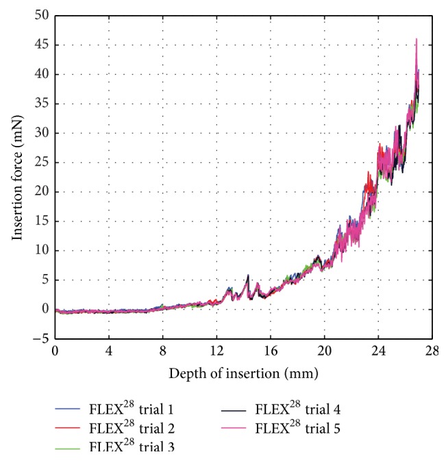
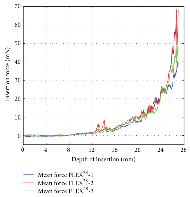
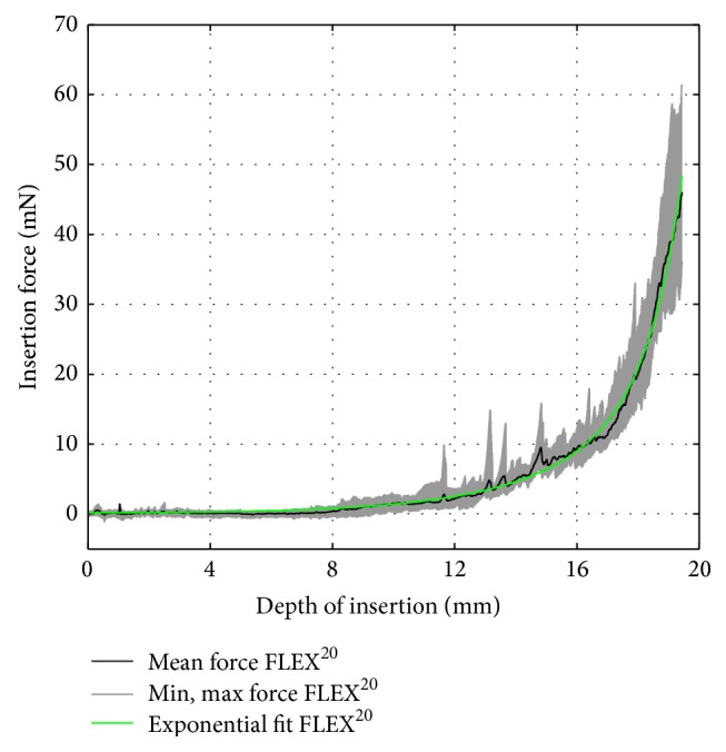
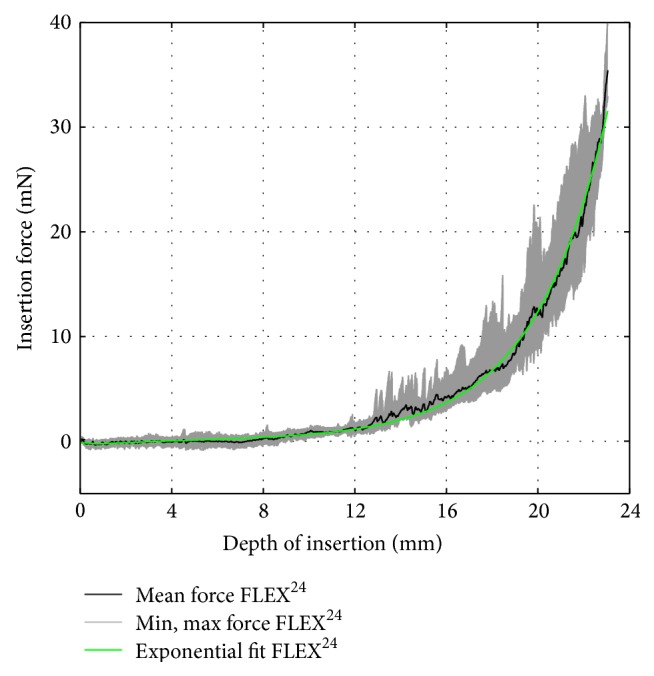
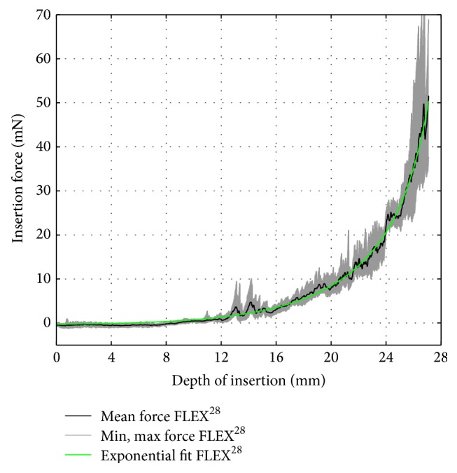
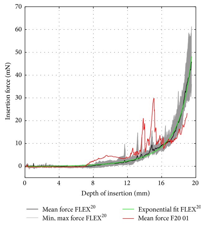
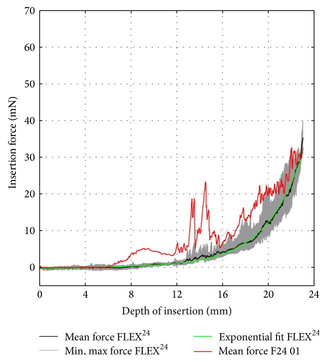
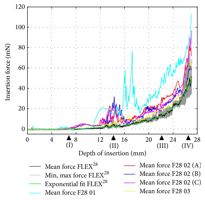
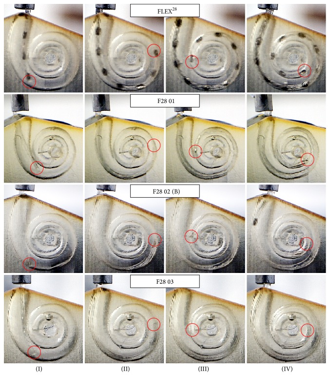
To develop skills sufficient for hearing preservation cochlear implant surgery, surgeons need to perform several electrode insertion trials in ex vivo temporal bones, thereby consuming relatively expensive electrode carriers. The objectives of this study were to evaluate the insertion characteristics of cochlear electrodes in a plastic scala tympani model and to fabricate radio opaque polymer filament dummy electrodes of equivalent mechanical properties. In addition, this study should aid the design and development of new cochlear electrodes. Automated insertion force measurement is a new technique to reproducibly analyze and evaluate the insertion dynamics and mechanical characteristics of an electrode. Mechanical properties of MED-EL's FLEX(28), FLEX(24), and FLEX(20) electrodes were assessed with the help of an automated insertion tool. Statistical analysis of the overall mechanical behavior of the electrodes and factors influencing the insertion force are discussed. Radio opaque dummy electrodes of comparable characteristics were fabricated based on insertion force measurements. The platinum-iridium wires were replaced by polymer filament to provide sufficient stiffness to the electrodes and to eradicate the metallic artifacts in X-ray and computed tomography (CT) images. These low-cost dummy electrodes are cheap alternatives for surgical training and for in vitro, ex vivo, and in vivo research purposes.
Figures
Similar articles
-
Automated insertion of preformed cochlear implant electrodes: evaluation of curling behaviour and insertion forces on an artificial cochlear model.Int J Comput Assist Radiol Surg. 2010 Mar;5(2):173-81. doi: 10.1007/s11548-009-0299-9. Epub 2009 Apr 10. Int J Comput Assist Radiol Surg. 2010. PMID: 20033522
-
Insertion depth impacts speech perception and hearing preservation for lateral wall electrodes.Laryngoscope. 2017 Oct;127(10):2352-2357. doi: 10.1002/lary.26467. Epub 2017 Mar 17. Laryngoscope. 2017. PMID: 28304096 Free PMC article.
-
Insertion Depth in Cochlear Implantation and Outcome in Residual Hearing and Vestibular Function.Ear Hear. 2016 Mar-Apr;37(2):e129-37. doi: 10.1097/AUD.0000000000000241. Ear Hear. 2016. PMID: 26524566
-
Potential benefits from deeply inserted cochlear implant electrodes.Ear Hear. 2011 Jul-Aug;32(4):411-27. doi: 10.1097/AUD.0b013e3182064bda. Ear Hear. 2011. PMID: 21248642 Review.
-
The rational for a mid-scala electrode array.Eur Ann Otorhinolaryngol Head Neck Dis. 2016 Jun;133 Suppl 1:S61-2. doi: 10.1016/j.anorl.2016.05.002. Epub 2016 May 27. Eur Ann Otorhinolaryngol Head Neck Dis. 2016. PMID: 27246747 Review.
Cited by
-
The role of pressure and friction forces in automated insertion of cochlear implants.Front Neurol. 2024 Aug 6;15:1430694. doi: 10.3389/fneur.2024.1430694. eCollection 2024. Front Neurol. 2024. PMID: 39170077 Free PMC article.
-
Investigation of ultra-low insertion speeds in an inelastic artificial cochlear model using custom-made cochlear implant electrodes.Eur Arch Otorhinolaryngol. 2018 Dec;275(12):2947-2956. doi: 10.1007/s00405-018-5159-1. Epub 2018 Oct 9. Eur Arch Otorhinolaryngol. 2018. PMID: 30302574
-
Evaluation of Real-Time Intracochlear Electrocochleography for Guiding Cochlear Implant Electrode Array Position.J Clin Med. 2023 Nov 29;12(23):7409. doi: 10.3390/jcm12237409. J Clin Med. 2023. PMID: 38068461 Free PMC article.
-
Robotic assistance during cochlear implantation: the rationale for consistent, controlled speed of electrode array insertion.Front Neurol. 2024 Jan 22;15:1335994. doi: 10.3389/fneur.2024.1335994. eCollection 2024. Front Neurol. 2024. PMID: 38318440 Free PMC article. Review.
-
Models of Cochlea Used in Cochlear Implant Research: A Review.Ann Biomed Eng. 2023 Jul;51(7):1390-1407. doi: 10.1007/s10439-023-03192-3. Epub 2023 Apr 22. Ann Biomed Eng. 2023. PMID: 37087541 Free PMC article. Review.
References
Publication types
MeSH terms
LinkOut - more resources
Full Text Sources
Other Literature Sources
Medical
