. 2015 Aug 6;10(8):e0129668.
doi: 10.1371/journal.pone.0129668. eCollection 2015.

Phylogeny of Echinoderm Hemoglobins

Affiliations

Phylogeny of Echinoderm Hemoglobins

Ana B Christensen et al. PLoS One. .

Abstract

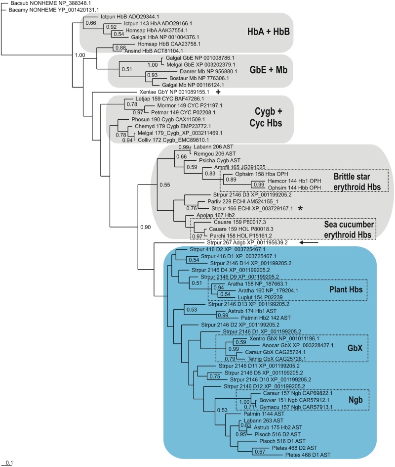
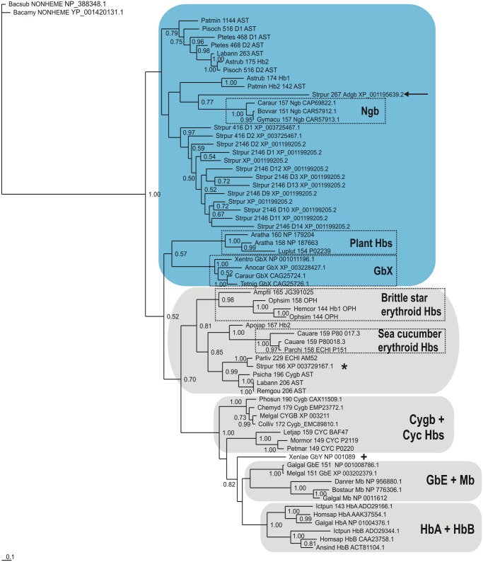
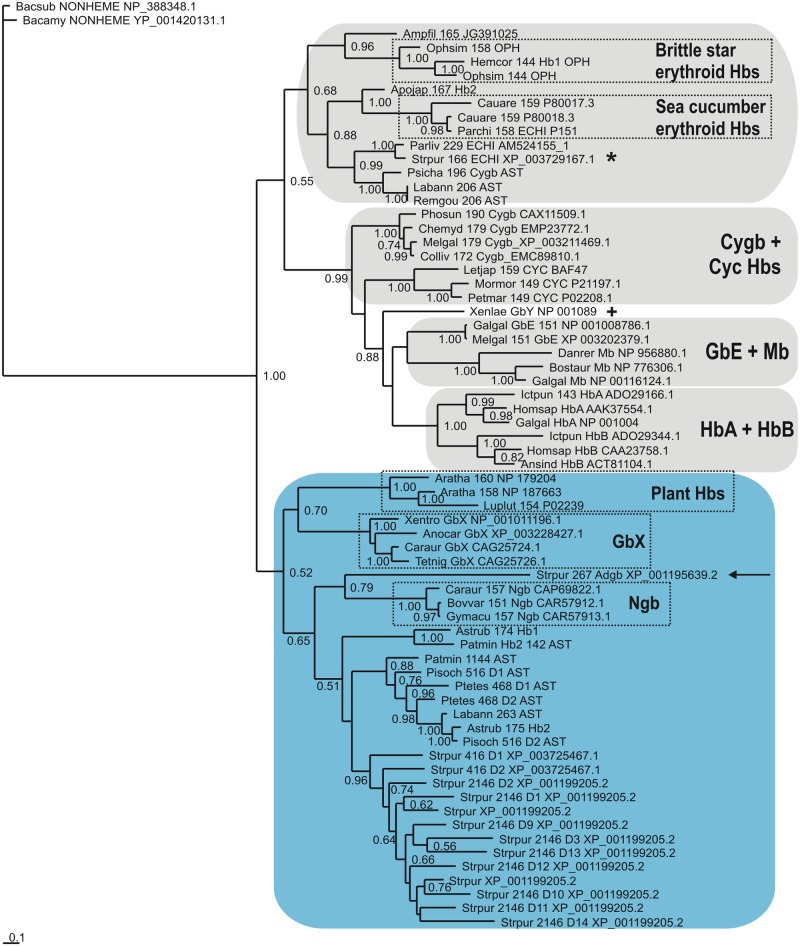
Background: Recent genomic information has revealed that neuroglobin and cytoglobin are the two principal lineages of vertebrate hemoglobins, with the latter encompassing the familiar myoglobin and α-globin/β-globin tetramer hemoglobin, and several minor groups. In contrast, very little is known about hemoglobins in echinoderms, a phylum of exclusively marine organisms closely related to vertebrates, beyond the presence of coelomic hemoglobins in sea cucumbers and brittle stars. We identified about 50 hemoglobins in sea urchin, starfish and sea cucumber genomes and transcriptomes, and used Bayesian inference to carry out a molecular phylogenetic analysis of their relationship to vertebrate sequences, specifically, to assess the hypothesis that the neuroglobin and cytoglobin lineages are also present in echinoderms.

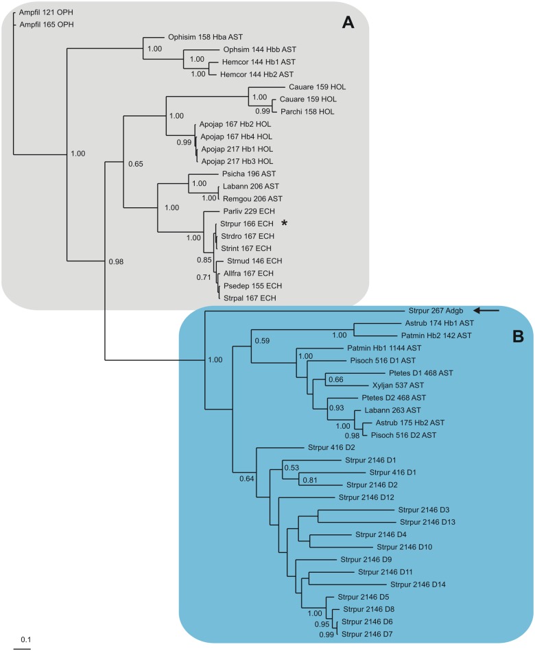
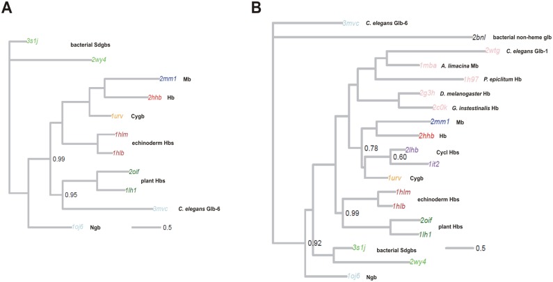
Results: The genome of the sea urchin Strongylocentrotus purpuratus encodes several hemoglobins, including a unique chimeric 14-domain globin, 2 androglobin isoforms and a unique single androglobin domain protein. Other strongylocentrotid genomes appear to have similar repertoires of globin genes. We carried out molecular phylogenetic analyses of 52 hemoglobins identified in sea urchin, brittle star and sea cucumber genomes and transcriptomes, using different multiple sequence alignment methods coupled with Bayesian and maximum likelihood approaches. The results demonstrate that there are two major globin lineages in echinoderms, which are related to the vertebrate neuroglobin and cytoglobin lineages. Furthermore, the brittle star and sea cucumber coelomic hemoglobins appear to have evolved independently from the cytoglobin lineage, similar to the evolution of erythroid oxygen binding globins in cyclostomes and vertebrates.

Conclusion: The presence of echinoderm globins related to the vertebrate neuroglobin and cytoglobin lineages suggests that the split between neuroglobins and cytoglobins occurred in the deuterostome ancestor shared by echinoderms and vertebrates.


Conflict of interest statement

Competing Interests: The authors have declared that no competing interests exist.

Figures

Fig 1
Fig 1. Bayesian phylogenetic tree based on a MAFFT L-INS-i MSA of 52 Echinoderm Hbs.
Fig 2
Fig 2. Bayesian phylogenetic tree based on a MAFFT L-INS-i MSA of 36 Echinoderm Hbs and 30 Vertebrate Hbs, using the Bacillus nonheme globin sequence [56], as outgroup.
Fig 3
Fig 3. Bayesian phylogenetic tree based on a T-Coffee Expresso MSA of 36 Echinoderm Hbs and 30 Vertebrate Hbs, using the Bacillus nonheme globin sequence [56], as outgroup.
Fig 4
Fig 4. Bayesian phylogenetic tree based on a Clustal Omega MSA of 36 Echinoderm Hbs and 30 Vertebrate Hbs, using the Bacillus nonheme globin sequence [56], as outgroup.
Fig 5
Fig 5. (A) Consensus tree generated by StructAlign, which carries out joint Bayesian inference of alignments and trees under a joint model of sequence and structure evolution.
The structures used here correspond to echinoderm coelomic Hbs (1hlb,1hlm), vertebrate Ngb (1oj6), Cygb (1urv), Mb (2mm1), HbA (2hhb) C. elegans Ngb (3mvc), plant Hbs (2oif, 1lh1), and two bacterial SDgbs (3s1j,2wy4). (B) Tree generated using a larger dataset, consisting of the aforementioned structures augmented with cyclostome Hbs (2lhb, 1it2) A. limacina Mb (1mba), D. melanogaster Hb (2g3h), G. intestinalis Hb (1c0k), C. elegans Glb-1 (2wtg), C. lacteus Ngb (2xki), and an bacterial non-heme globin (1bnl) as an outgroup.
Fig 6
Fig 6. Structural superposition of the echinoderm structure 1hlm (brown) with the cytoglobin structure 1urv (blue).
The two structures show a very close correspondence, with some localized deviations, such as at helix D, which is disordered in 1hlm (marked by Arg65), and at the C-terminal end of helix G (marked by Val131).
Fig 7
Fig 7. Analysis of average per-site root mean square deviation (RMSD) between the echinoderm structures (1hlb, 1hlm) and the Ngb (1oj6) and Cygb (1urv) structures.
Structural deviation was computed from each of the echinoderms to the target structure of interest as described in the Methods section, using the consensus alignment generated on the larger structural dataset; the mean of these two values computed for each column. Colored blocks at the bottom indicate charged (red) and non-charged (black) residues. Gaps are shown in grey, and the proximal and distal histidines are shown in blue. Helix locations are annotated above, and named using standard conventions. Overall RMSD for each plot is computed as the mean of the squared contributions from each site, and is indicated by the dashed red line. The blue areas underneath indicate the confidence associated with each column in the multiple alignment, as outputted by StructAlign.
Fig 8
Fig 8. Analysis of pairwise per-site root mean square deviation (RMSD) between the echinoderm structures (1hlb, 1hlm) and the Ngb (1oj6) and Cygb (1urv) structures.
Colored blocks at the bottom indicate charged (red) and non-charged (black) residues. Gaps are shown in grey, and the proximal and distal histidines are shown in blue. Helix locations are annotated above, and names using standard conventions. Overall RMSD for each plot is computed as the mean of the squared contributions from each site, and is indicated by the dashed red line. The blue areas underneath indicate the confidence associated with each column in the multiple alignment, as outputted by StructAlign.

References

    1. Vinogradov SN, Hoogewijs D, Bailly X, Arredondo-Peter R, Guertin M, Gough J, et al. Three globin lineages belonging to two structural classes in genomes from the three kingdoms of life. Proc Natl Acad Sci U S A. 2005;102(32):11385–9. - PMC - PubMed
    1. Burmester T, Weich B, Reinhardt S, Hankeln T. A vertebrate globin expressed in the brain. Nature. 2000;407(6803):520–3. - PubMed
    1. Burmester T, Ebner B, Weich B, Hankeln T. Cytoglobin: a novel globin type ubiquitously expressed in vertebrate tissues. Mol Biol Evol. 2002;19(4):416–21. 11919282 - PubMed
    1. Kawada N, Kristensen DB, Asahina K, Nakatani K, Minamiyama Y, Seki S, et al. Characterization of a stellate cell activation-associated protein (STAP) with peroxidase activity found in rat hepatic stellate cells. J Biol Chem. 2001;276(27):25318–23. - PubMed
    1. Trent JT 3rd, Hargrove MS. A ubiquitously expressed human hexacoordinate hemoglobin. J Biol Chem. 2002;277(22):19538–45. - PubMed

Publication types