. 2015 Aug 6;11(8):e1005429.
doi: 10.1371/journal.pgen.1005429. eCollection 2015 Aug.

A Genome Scale Screen for Mutants with Delayed Exit from Mitosis: Ire1-Independent Induction of Autophagy Integrates ER Homeostasis into Mitotic Lifespan

Affiliations

A Genome Scale Screen for Mutants with Delayed Exit from Mitosis: Ire1-Independent Induction of Autophagy Integrates ER Homeostasis into Mitotic Lifespan

Ata Ghavidel et al. PLoS Genet. .

Abstract

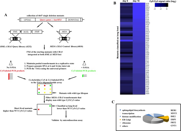
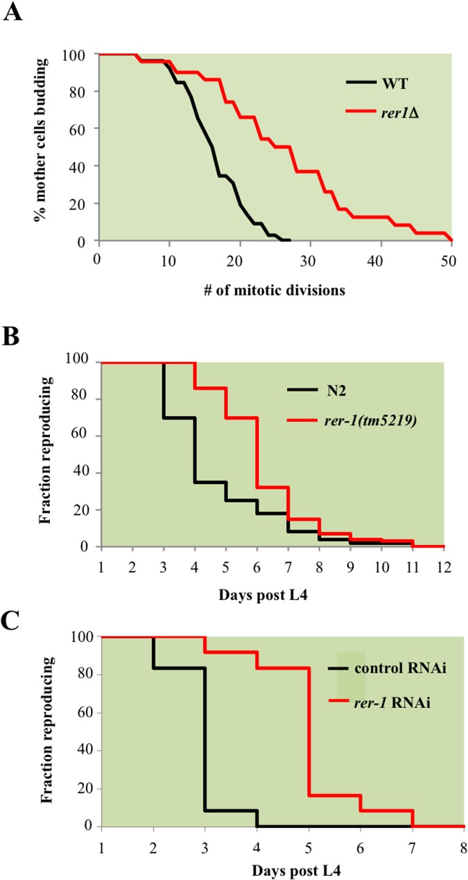
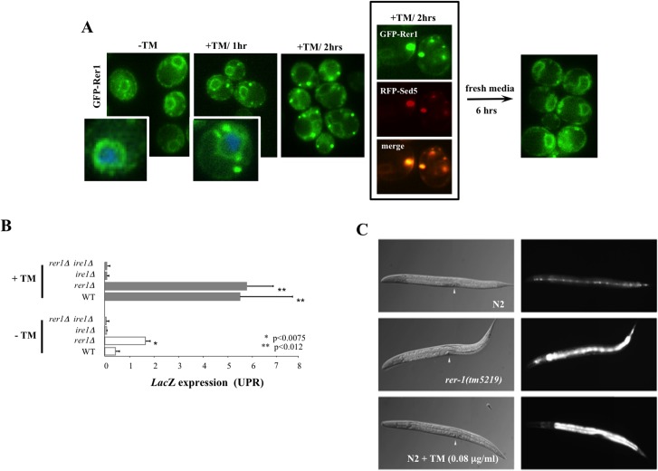
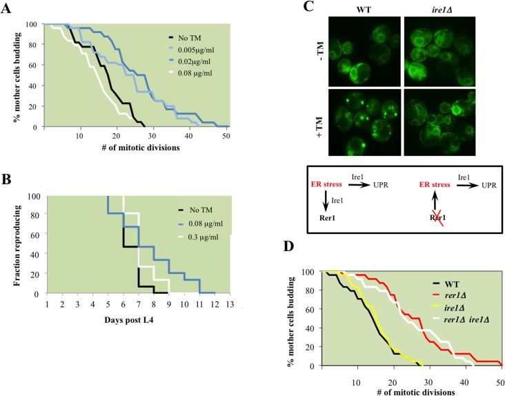
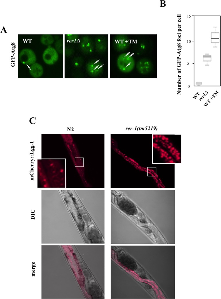
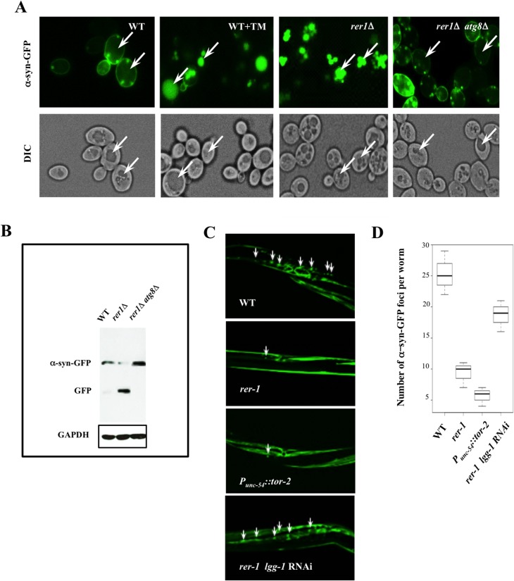
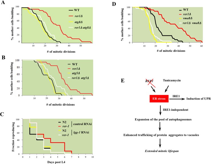
Proliferating eukaryotic cells undergo a finite number of cell divisions before irreversibly exiting mitosis. Yet pathways that normally limit the number of cell divisions remain poorly characterized. Here we describe a screen of a collection of 3762 single gene mutants in the yeast Saccharomyces cerevisiae, accounting for 2/3 of annotated yeast ORFs, to search for mutants that undergo an atypically high number of cell divisions. Many of the potential longevity genes map to cellular processes not previously implicated in mitotic senescence, suggesting that regulatory mechanisms governing mitotic exit may be broader than currently anticipated. We focused on an ER-Golgi gene cluster isolated in this screen to determine how these ubiquitous organelles integrate into mitotic longevity. We report that a chronic increase in ER protein load signals an expansion in the assembly of autophagosomes in an Ire1-independent manner, accelerates trafficking of high molecular weight protein aggregates from the cytoplasm to the vacuoles, and leads to a profound enhancement of daughter cell production. We demonstrate that this catabolic network is evolutionarily conserved, as it also extends reproductive lifespan in the nematode Caenorhabditis elegans. Our data provide evidence that catabolism of protein aggregates, a natural byproduct of high protein synthesis and turn over in dividing cells, is among the drivers of mitotic longevity in eukaryotes.


Conflict of interest statement

The authors have declared that no competing interests exist.

Figures

Fig 1
Fig 1. A genome scale screen for isolating mutants with extended mitotic lifespan in the yeast S. cerevisiae.
A. The screen work flow. The design rationale is detailed in S1 Fig. B. Cy5:Cy3 signal ratios of mutants maintained in a dividing state for 16 days. Ranked values were log2 normalized and projected. Of the starting collection of 3762, 52 mutants that maintained negative log2 Cy5:Cy3 signal ratios at both day 6 and day 16 and displayed signal ratios <-2.3 at day 16 were classified as potentially long-lived (S3 Fig). C. Broad functional clustering of the putative longevity genes isolated in this screen using GO Ontology. Genes that function in protein modification and trafficking across the ER-Golgi network are outlined.
Fig 2
Fig 2. RER1 inactivation extends mitotic lifespan in yeast and reproductive lifespan in C. elegans.
A. Mitotic lifespan of wild type yeast and an isogenic rer1Δ grown in complete media (YPD) at 30°C. Buds from a starting population of 40 founding mother cell were successively dissected and counted until all mother cells had ceased dividing. The mean mitotic lifespan for WT and rer1∆ was 17.5 and 27.5 days, respectively. B. Representative reproductive lifespan of self fertilized wild type (n = 85) and rer-1(tm5129) (n = 84) hermaphrodites. Fraction of adults generating viable progeny as a function of time is plotted. Day 1 corresponds to the first egg-laying day of adulthood (around 9 hours after the L4 lethargus at 20°C). C. Representative reproductive lifespan analysis in worms treated with control (empty vector) or rer-1 RNAi. In comparison to OP50 supplemented plates (B), growth on RNAi plates supplemented with HT115 E. Coli routinely accelerates the rate of progeny production where on average by the end of day 3 wild type hermaphrodites had generated nearly 90% of their total progeny [48].
Fig 3
Fig 3. Induction of UPR in yeast and worm rer1 mutants.
A. Live cell imaging of GFP-Rer1 expressed from a centromeric plasmid in yeast rer1Δ mutants treated with DMSO or after exposure 0.2 μg/ml TM. GFP-Rer1 localizes to perinuclear ER in untreated cells and redistributes to Golgi in cells treated with TM. Insets show cells counterstained with DAPI. Boxed images denote GFP-Rer1 colocalization with Golgi resident RFP-Sed5. GFP-Rer1 relocalizes to ER following a 6 hour release into fresh media. B. The in situ expression of LacZ from a KAR2 promoter after a 2 hour exposure to DMSO or 0.2 μg/ml TM. Data represent means ± s.e.m. (n = 3). C. hsp-4::gfp expression in wild type, rer-1(tm5219) or TM-exposed adult hermaphrodites at 20°C. Background fluorescence in untreated N2 is due to basal expression of the GFP reporter in seam cells. Arrowheads denote vulva.
Fig 4
Fig 4. ER stress extends reproductive lifespan in yeast and worms.
A. Mitotic lifespan of wild type yeast grown on YPD supplemented with the indicated concentrations of TM. Mean mitotic lifespans for WT (18.5) and cells treated with TM at 0.005 (24.5) and 0.02 (27.5) μg/ml were statistically significant (p-values<0.01). B. Reproductive lifespan in worms with or without dietary supplementation with TM. C. Live cell imaging of GFP-Rer1 in wild type yeast and ire1Δ mutants treated with DMSO or after a 2 hr exposure to 0.02 μg/ml TM. (Box) In tandem with inducing canonical UPR, ER stress increases trafficking from ER to Golgi, reflected in the Golgi redistribution of GFP-Rer1 in cells treated with TM. Conversely, deleting RER1 increases ER load denoted by the high basal UPR in rer1Δ mutants. D. Mitotic lifespan of WT (16.5 days) and isogenic rer1∆ (26.5), ire1∆ (16.5) and rer1∆ ire1∆ (24.5) mutants at 30°C. Differences between WT mean mitotic lifespan were significant for rer1∆ and rer1∆ ire1∆ mutants (p-values<0.001).
Fig 5
Fig 5. Induction of autophagy in yeast and worm rer1 mutants.
A. Live cell imaging of GFP-Atg8 foci (arrows) in wild type cells after a 2 hr exposure to TM (0.02 μg/ml) and rer1Δ cells. B. Microscopically detectable GFP foci in individual yeast cells were scored from digitized Z-stacked images by an observer blind to the genotype. Data represent means ± s.e.m. (n>20). C. Intestinal expression of mCherry-LGG-1 (P nhx-2 mCherry::lgg- 1) in day2 wild type and rer-1(tm5219) hermaphrodites. Insets represent magnification of the boxed sections.
Fig 6
Fig 6. Enhanced clearance of protein aggregates in yeast and worm rer1 mutants.
A. Live cell imaging of α-syn-GFP after a 4 hr induction in 0.2% galactose. TM (0.02 μg/ml) was added to the cultures after an initial induction in galactose and cells were imaged 2 hours later. In a marked contrast to untreated cells, the GFP foci were largely vacuolar in rer1Δ or wild type cells treated with TM (arrows). B. GFP immunoblot in whole cell lysates prepared from cells in (A). The intravacuolar proteolysis of α-syn-GFP generates the transiently stable GFP moiety detected as a discrete fragment in SDS-PAGE. C. Fluorescent images of transgenic hermaphrodites stably expressing α-syn-GFP from a body wall muscle promoter (Punc- 54::α-syn::GFP). Arrows denote a subset of the α-syn-GFP foci. Worms co-expressing heat shock protein torsinA (tor-2) in the same cells (P unc-54::tor-2) served as a positive experimental control. The average number of α-syn-GFP foci per worm scored from digitized Z-stacked images ± s.e.m are plotted in D (n >20).
Fig 7
Fig 7. The requirement for intact cellular catabolism in extension of lifespan in yeast and worm rer1 mutants.
Mitotic lifespan in wild type or rer1Δ mutants harboring a deletion of the autophagosome components ATG8 (A) or ATG5 (B). The mean mitotic lifespans for the samples in (A) were WT (16.5 days), rer1∆ (27.5), atg8∆ (15.5) and rer1∆ atg8∆ (18.5). The rer1∆ mean mitotic lifespan was significantly different from the WT (p-value<0.01), whereas the differences between WT, atg8∆ and rer1∆ atg8∆ were not. Similarly, in (B) the mean mitotic lifespan difference between WT (17.5 days) and rer1∆ (38.5) was statistically significant (p-value<0.01), whereas the differences between WT, atg5∆ (17.5), and rer1∆ atg5∆ (18.5) were not. C. Reproductive lifespan in N2 (n = 25) and rer-1(tm5219) (n = 25) worms treated with control or lgg-1 RNAi. D. Deletion of VMA8, encoding a vacuolar proton pump subunit, abolishes the extended lifespan in yeast rer1Δ mutants. The differences between WT (21.5 days), vma8∆ (15.5; p-value < 0.01) and rer1∆ vma8∆ (17.5; p-value < 0.05) were statistically significant. E. Autophagy, induced in response to elevated ER stress in rer1Δ or following exposure to the ER stressor (TM), traffics cytoplasmic aggregates to the vacuoles and in doing so delays mitotic senescence (details in text).

References

    1. Hayflick L (1965) The Limited in Vitro Lifetime of Human Diploid Cell Strains. Exp Cell Res 37: 614–636. - PubMed
    1. Smith JR, Pereira-Smith OM (1996) Replicative senescence: implications for in vivo aging and tumor suppression. Science 273: 63–67. - PubMed
    1. Jazwinski SM, Egilmez NK, Chen JB (1989) Replication control and cellular life span. Exp Gerontol 24: 423–436. - PubMed
    1. Kaeberlein M, Powers RW 3rd, Steffen KK, Westman EA, Hu D, et al. (2005) Regulation of yeast replicative life span by TOR and Sch9 in response to nutrients. Science 310: 1193–1196. - PubMed
    1. Lesur I, Campbell JL (2004) The transcriptome of prematurely aging yeast cells is similar to that of telomerase-deficient cells. Mol Biol Cell 15: 1297–1312. - PMC - PubMed

Publication types

MeSH terms

Substances