Review
. 2015 Aug;21(8):854-62.
doi: 10.1038/nm.3918.

The central role of muscle stem cells in regenerative failure with aging

Affiliations
Review

The central role of muscle stem cells in regenerative failure with aging

Helen M Blau et al. Nat Med. 2015 Aug.

Abstract

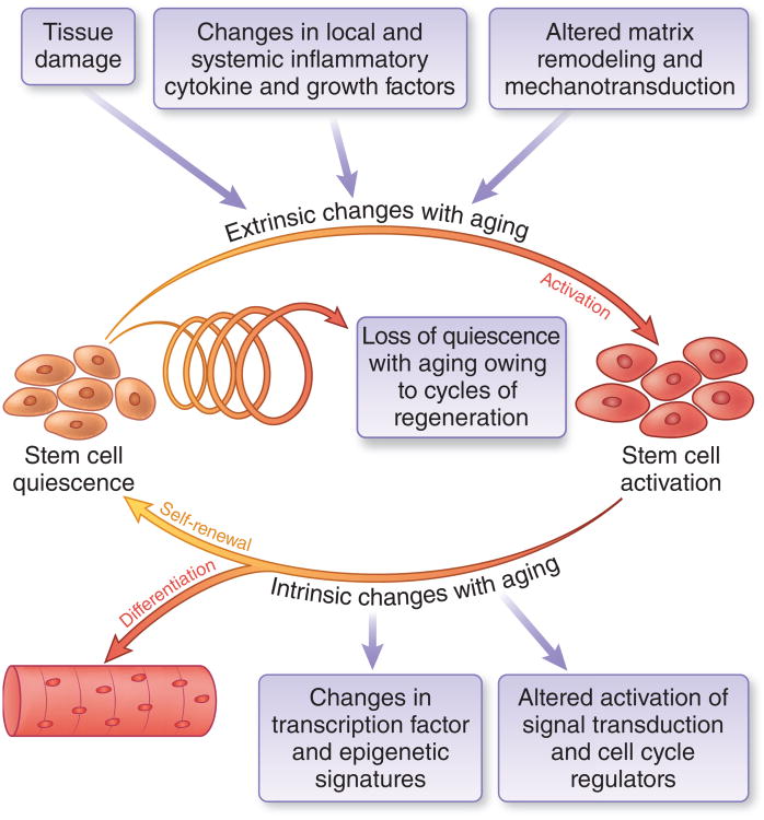
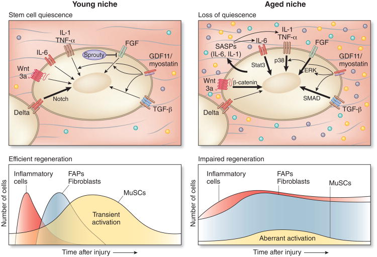
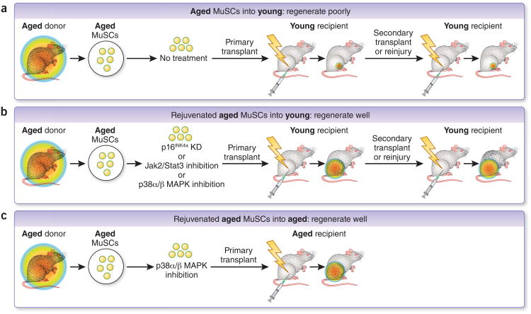
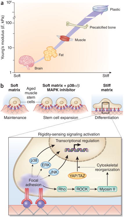
Skeletal muscle mass, function, and repair capacity all progressively decline with aging, restricting mobility, voluntary function, and quality of life. Skeletal muscle repair is facilitated by a population of dedicated muscle stem cells (MuSCs), also known as satellite cells, that reside in anatomically defined niches within muscle tissues. In adult tissues, MuSCs are retained in a quiescent state until they are primed to regenerate damaged muscle through cycles of self-renewal divisions. With aging, muscle tissue homeostasis is progressively disrupted and the ability of MuSCs to repair injured muscle markedly declines. Until recently, this decline has been largely attributed to extrinsic age-related alterations in the microenvironment to which MuSCs are exposed. However, as highlighted in this Perspective, recent reports show that MuSCs also progressively undergo cell-intrinsic alterations that profoundly affect stem cell regenerative function with aging. A more comprehensive understanding of the interplay of stem cell-intrinsic and extrinsic factors will set the stage for improving cell therapies capable of restoring tissue homeostasis and enhancing muscle repair in the aged.


Figures

Figure 1
Figure 1
The role of MuSCS in tissue homeostasis with aging. In adult muscles, MuSCs are maintained in quiescence. During muscle regeneration, MuSCs are transiently activated and self-renew to produce more stem cells and differentiated progeny, maintaining tissue homeostasis and repair capacity. Repair is initiated by tissue damage followed by a two-phase inflammatory response involving secretion of pro- and antiinflammatory cytokines. ‘Extrinsic’ microenvironmental factors governing this process are provided by neighboring cells and from systemic sources. Cytokines trigger the production of matrix degrading enzymes leading to extracellular matrix remodeling. These extrinsic stimuli converge to trigger ntrinsic changes in MuSCs signaling, cell cycle, and transcriptional networks that regulate self-renewal and differentiation. With aging, progressive cycles of damage and repair lead to a loss of homeostasis, resulting in depletion of quiescent MuSCs with self-renewal capacity, owing to changes in both extrinsic and instrinic factors, resulting in impaired muscle regeneration.
Figure 2
Figure 2
Extrinsic and intrinsic regulators of MuSC function are altered in aging. In young muscles, MuSCs are transiently activated after tissue damage by released inflammatory cytokines and growth factor signaling coordinated by immune cells, fibroblasts and FAPs. These factors dynamically stimulate MuSC regulatory pathways, including Delta-Notch, governing cell cycle and transcription control of MuSC fate, resulting in temporary activation and self-renewal, with homeostasis reached upon completion of myofiber repair through re-induction of stem cell quiescence, in part via activation of Sprouty, an inhibitor of FGF signaling,. The thickness of the arrow indicates strength of activation of the pathway in young and aged MuSCs. In aged muscles, self-renewal signals (for example, Delta) diminish and inflammatory and fibrogenic signals are elevated and prolonged,, resulting in aberrant MuSC activation and loss of quiescence,,. These changes with aging are commensurate with the onset of aberrant activation in the β-catenin, Stat3, FGF, p38α/β MAPK, and SMAD signaling pathways. By elderly ages, a subset of aged MuSCs become senescent,.
Figure 3
Figure 3
Evidence in support of intrinsic ‘memory’ in aged and rejuvenated MuSC populations. (a) Rainbow colored circles represent the ability to regenerate. A heritable cell-intrinsic defect in regenerative capacity of aged MuSCs is evident upon serial transplantation into young recipients. Regenerative capacity remains poor even in a young tissue microenvironment,,,. (b) After treatment with various ex vivo ‘rejuvenating’ treatments perturbing p16Ink4a, Jak2-Stat3, or p38α/β MAPK activity and transplantation into a young muscle microenvironment,,, aged MuSC cells are capable of efficient regeneration even in serial transplantation. Factors intrinsic and extrinsic (i.e., in the recipient microenvironment) to the rejuvenated aged MuSCs may corroborate to enhance regenerative capacity in this setting,,. (c) Rejuvenation of aged muscle stem cells after ex vivo exposure to soft hydrogel substrate and a chemical p38α/β MAPK inhibitor co-treatment leads to robust transplantation even in an aged tissue microenvironment, leading to enhanced muscle strength.
Figure 4
Figure 4
Biophysical regulation of muscle stem cells in aging. (a) Tissue elasticity, the biophysical property of tissues and cell culture materials most commonly associated with effects on cell proliferation and differentiation, both in stem cells from skeletal muscle and other tissues, is typically characterized by a Young's modulus (E). The Young's modulus describes the elastic deformability of a material, and is proportional to the amount of force (stress) required to achieve a given deformation (strain). Characteristic tissue elasticities include brain and mammary glands (E = ∼1 kPa), adipose (∼5 kPa), skeletal muscle (∼10−30 kPa, and precalcified bone (∼50 kPa), Tissue culture substrates such as polystyrene and glass have typical Young's moduli of ∼106 kPa (ref. 114). In aging and muscular dystrophy, skeletal muscles become progressively fibrotic, resulting in increased rigidity,. (b) For muscle stem cells from aged mice, a soft 12-kPa matrix (mimicking the rigidity of healthy skeletal muscle) supports muscle stem cell self-renewal expansion in synergy with targeted inhibition of the p38α/β MAPK signaling pathway. A soft matrix alone supports aged MuSC maintenance whereas a stiff (106 kPa) matrix induces the differentiation of aged MuSCs. Cells sense the rigidity of their surrounding substrate through a variety of adhesion-dependent signaling pathways, which result in cellular deformations (for example, elongation) and transcriptional responses. Across many cell types, substrate rigidity is sensed by integrin clustering and focal adhesion stabilization, which yields prolonged activation of both canonica MAPK pathways (p38, ERK, JNK) and Rho/Rho-associated kinase (ROCK) signaling and subsequent myosin II–mediated cytoskeletal reorganization, leading to YAP/TAZ transcription factor translocalization. Together, these and other adhesion-activated signaling pathways confer sensitivity to matrix stiffness in numerous cell types.

References

    1. Buchman TG. The community of the self. Nature. 2002;420:246–251. - PubMed
    1. Seale P, Asakura A, Rudnicki MA. The potential of muscle stem cells. Dev Cell. 2001;1:333–342. - PubMed
    1. Cheung TH, Rando TA. Molecular regulation of stem cell quiescence. Nat Rev Mol Cell Biol. 2013;14:329–340. - PMC - PubMed
    1. Montarras D, L'Honore A, Buckingham M. Lying low but ready for action: the quiescent muscle satellite cell. FEBS J. 2013;280:4036–4050. - PubMed
    1. Bosnakovski D, et al. Prospective isolation of skeletal muscle stem cells with a Pax7 reporter. Stem Cells. 2008;26:3194–3204. - PMC - PubMed

Publication types

Substances