. 2015 Oct;59(10):6210-22.
doi: 10.1128/AAC.01406-15. Epub 2015 Jul 27.

Key Residues of Outer Membrane Protein OprI Involved in Hexamer Formation and Bacterial Susceptibility to Cationic Antimicrobial Peptides

Affiliations

Key Residues of Outer Membrane Protein OprI Involved in Hexamer Formation and Bacterial Susceptibility to Cationic Antimicrobial Peptides

Ting-Wei Chang et al. Antimicrob Agents Chemother. 2015 Oct.

Abstract

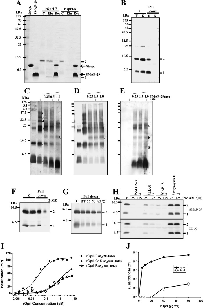
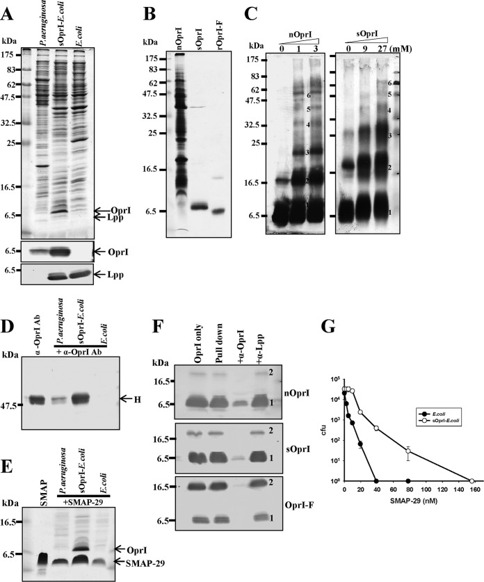
Antimicrobial peptides (AMPs) are important components of the host innate defense mechanism against invading pathogens. Our previous studies have shown that the outer membrane protein, OprI from Pseudomonas aeruginosa or its homologue, plays a vital role in the susceptibility of Gram-negative bacteria to cationic α-helical AMPs (Y. M. Lin, S. J. Wu, T. W. Chang, C. F. Wang, C. S. Suen, M. J. Hwang, M. D. Chang, Y. T. Chen, Y. D. Liao, J Biol Chem 285:8985-8994, 2010, http://dx.doi.org/10.1074/jbc.M109.078725; T. W. Chang, Y. M. Lin, C. F. Wang, Y. D. Liao, J Biol Chem 287:418-428, 2012, http://dx.doi.org/10.1074/jbc.M111.290361). Here, we obtained two forms of recombinant OprI: rOprI-F, a hexamer composed of three disulfide-bridged dimers, was active in AMP binding, while rOprI-R, a trimer, was not. All the subunits predominantly consisted of α-helices and exhibited rigid structures with a melting point centered around 76°C. Interestingly, OprI tagged with Escherichia coli signal peptide was expressed in a hexamer, which was anchored on the surface of E. coli, possibly through lipid acids added at the N terminus of OprI and involved in the binding and susceptibility to AMP as native P. aeruginosa OprI. Deletion and mutation studies showed that Cys1 and Asp27 played a key role in hexamer formation and AMP binding, respectively. The increase of OprI hydrophobicity upon AMP binding revealed that it undergoes conformational changes for membrane fusion. Our results showed that OprI on bacterial surfaces is responsible for the recruitment and susceptibility to amphipathic α-helical AMPs and may be used to screen antimicrobials.


Figures

FIG 1
FIG 1
Preparation and configuration of two recombinant OprIs, rOprI-F and rOprI-R. (A) Analysis of recombinant OprI at different purification steps. Proteins were separated by 14% SDS-PAGE and stained by Coomassie blue. Lane 1, crude lysate from E. coli BL21(DE3) transformed with a maltose-binding protein (MBP)/OprI-fused gene; lane 2, eluate of nickel-affinity gel; lane 3, protease factor Xa-digested product; lanes 4 and 5, supernatant and pellet of protease-digested products after heat treatment and centrifugation; lanes 6 and 7, front peak (rOprI-F) and rear peak (rOprI-R) of Superose 12 column eluates; lanes 8 and 9, rOprI-F and rOprI-R analyzed by nonreducing SDS-PAGE. (B) Gel filtration chromatography of rOprI. Immunoglobulin (IgG; 150 kDa), bovine serum albumin (BSA; 66 kDa), and bovine RNase A (RNase A; 14 kDa) were used as molecular size markers. (C) SEC-MALS analysis of rOprI. Horizontal line segments above the elution peaks correspond to calculated molar mass (left ordinate axis), and the corresponding molecular mass is indicated in Daltons. The solid line corresponds to relative UV absorbance at 280 nm, and the dashed line in gray represents light scattering (right ordinate axis). (D and E) Configuration of rOprI-F and rOprI-R after cross-linking with EDC. rOprI-F and rOprI-R were incubated with increasing amounts of EDC, as indicated, at pH 5.5 for 30 min and subjected to 14% SDS-PAGE and Coomassie blue staining (4 μg protein in each lane) (D) and Western blot analysis (0.8 μg protein in each lane) (E). Numbers 1 to 6 represent the status of rOprI polymerization.
FIG 2
FIG 2
Circular dichroism (CD) spectrum analyses of OprI. (A) Circular dichroism spectrum analysis of 20 μM rOprI-F and rOprI-R in 20 mM HEPES, pH 7.4, 50 mM NaCl. M.E., molar ellipticity. (B) Equilibrium circular dichroism titration experiments of 20 μM rOprI-F and rOprI-R as a function of temperature monitored at 220 nm.
FIG 3
FIG 3
Differential AMP-binding ability of rOprI-F and rOprI-R. (A) Pulldown experiments by biotinylated SMAP-29-streptavidin gel. The biotinylated SMAP-29 was incubated with streptavidin-conjugated gels (Strep.) in 10 mM sodium phosphate, pH 7.5, and then with rOprI-F and rOprI-R (4 μg each), and finally it was eluted by 0.75% sodium N-dodecanoylsarcosinate, separated by nonreducing SDS-PAGE, and visualized by Coomassie blue staining. C, Elu, and Res represent control without pulldown, N-dodecanoylsarcosinate eluate, and residual proteins, respectively. (B) Pulldown experiments similar to those shown in panel A. The biotinylated SMAP-29 was incubated with streptavidin-conjugated beads in 10 mM sodium phosphate, pH 7.5, 0.15 M NaCl, and 0.15% Triton X-100, with rOprI-F and rOprI-R (2 μg each), and then separated by reduced SDS-PAGE and visualized by Western blotting using anti-OprI antibody. C, F, and R represent control, rOprI-F, and rOprI-R, respectively. (C to E) Formation of rOprI-F and SMAP-29 complex. rOprI-F (0.4 μg each) was incubated with increasing amounts of SMAP-29 for 30 min, cross-linked with 0.02% glutaraldehyde for 20 min, separated by 14% reduced SDS-PAGE, and analyzed by silver staining (C), Western blotting with anti-OprI antibody (D), or anti-SMAP-29 antibody (E). Numbers 1 to 6 represent the status of rOprI-F polymerization, and letters a to c indicate the positions of rOprI-F/SMAP-29 complexes. (F) Sensitivity of rOprI-F to reducing agent. rOprI-F was incubated in 15 mM 2-mercaptoethanol for 10 min at room temperature (RT) before SMAP-29-binding experiments as described for panel B. (G) Thermal stability of rOprI-F to bind SMAP-29. rOprI-F was treated at 55°C, 70°C, or 85°C for 10 min before binding to SMAP-29. (H) Competition of the formation of OprI-F-SMAP-29 complex by free cationic α-helical AMPs. rOprI-F (2 μg each) was incubated with biotinylated SMAP-29 in the presence of free SMAP-29, CAP-18, LL-37, and polymyxin B. (I) Fluorescence polarization of the interaction between FITC-SMAP-29 and various OprI variants. (J) Repression of antimicrobial activity of SMAP-29 by exogenous recombinant OprI. P. aeruginosa (5 × 10⁴ to 10 × 104 CFU) was treated with 0.1 μM SMAP-29 in the presence of exogenous rOprI-F and rOprI-R as indicated. At least three independent experiments were performed for each bactericidal assay to present average values with standard deviations. Some statistical bars could not be drawn clearly due to the very minor experimental variations at high CFU values.
FIG 4
FIG 4
Hexameric OprI expressed on the surface of E. coli having AMP-binding ability. (A) Expression of OprI. The total lysates of P. aeruginosa, sOprI-transformed E. coli RST-2, and parental cells (107 CFU) were separated by 14% SDS-PAGE and analyzed by Coomassie blue staining (top), Western blotting with anti-OprI antibody (middle), or anti-Lpp antibody (bottom), respectively. (B) SDS-PAGE analyses of OprI extracted from P. aeruginosa (nOprI; 21 μg), sOprI-transformed E. coli (sOprI; 2 μg), and recombinant OprI excised from MBP-fused protein (rOprI-F; 1.6 μg) visualized by Coomassie blue staining. (C) Analyses of oligomerization of nOprI (left) and sOprI (right). nOprI (0.7 μg each) and sOprI (0.02 μg each) were incubated with increasing amounts of EDC at pH 5.5, as indicated, containing 0.25% sodium N-dodecanoylsarcosinate for 30 min and subjected to 14% SDS-PAGE and Western blotting. (D and E) The binding abilities of P. aeruginosa, sOprI-transformed E. coli RST2, and parental cells to anti-OprI antibody (Ab) or SMAP-29. Bacteria (107 CFU) were incubated with anti-OprI antibody (4 μg) or SAMP-29 (3 μg) in 100 μl at 37°C for 30 min, washed by 0.4 M NaCl, and subjected to SDS-PAGE and Coomassie blue staining. H represents the heavy chain of immunoglobulin. (F) SMAP-29 binding ability of various OprIs. The biotinylated SMAP-29 bound to streptavidin-conjugated gels was incubated with nOprI (14 μg), sOprI (2 μg), and rOprI-F (2 μg), respectively, in 10 mM sodium phosphate, pH 7.5, 0.15 M NaCl, and 0.15% Triton X-100 in the absence or presence of anti-OprI and anti-Lpp antibodies (4 μg each) for blocking the AMP-binding activity of OprI. After pulling down and washing, the OprIs were visualized by Western blotting using anti-OprI antibody. (G) Susceptibilities of sOprI-transformed and parental E. coli RST2 cells to SMAP-29. The cold-stored bacteria (5 × 10⁴ to 10 × 104 CFU) were treated with SMAP-29 and assayed for their viabilities. Average values with standard deviations are presented.
FIG 5
FIG 5
Analyses of secondary structure and configurations of OprI variants. (A and C) Circular dichroism spectrum analyses of 20 μM OprI variants in 20 mM HEPES, pH 7.4, 50 mM NaCl. (B and D) Equilibrium circular dichroism titration experiments as a function of temperature monitored at 220 nm. (E to G) EDC cross-linking of OprI variants. The OprI variants (0.8 μg each) were incubated with 125 mM EDC at pH 5.5 for 30 min and subjected to 14% SDS-PAGE and Western blotting. Numbers 1 to 8 represent the status of rOprI polymerization.
FIG 6
FIG 6
AMP-binding abilities of OprI variants. (A, bottom) The OprI variants (2 μg each) were pulled downed by biotinylated SMAP-29 as described in the legend to Fig. 3B. (Top) A volume of 0.4 μg of each rOprI variant was used as a control without the pulldown experiment. (B, C, and D) Protection of bacteria from AMP action by OprI variants. The viability of P. aeruginosa (5 × 10⁴ to 10 × 104 CFU) was determined after being treated with 0.1 μM SMAP-29 in the presence of OprI variants. Average values with standard deviations are presented.
FIG 7
FIG 7
Changes of emission spectra of OprI-associated ANS driven by SMAP-29. The emission spectra of ANS in the presence of 10 μg/ml SMAP-29 and/or 80 μg/ml rOprI were measured in 200 μl solution. ANS was added and adjusted to the indicated concentrations (lines 1 to 6 at 0, 5, 10, 20, 30, and 40 μM, respectively). Arrows indicate the wavelength of emission maximum at 470 nm or 520 nm, emitted by bound- and free-form ANS, respectively. (A) ANS alone; (B) SMAP-29; (C) rOprI-F; (D) rOprI-F plus SMAP-29; (E) rOprI-R; (F) rOprI-R plus SMAP-29.
FIG 8
FIG 8
Proposed model of the structure of trimeric and hexameric OprIs. (A) Helical wheel projections of residues 10 to 64 of monomeric OprI. The 4,3 hydrophobic heptad repeats are labeled in a through g positions. (B) Three-stranded α-helices showing hydrophobic interactions between alanine residues in positions a and d. (C) Diagram of hexameric OprI and AMP complex. Three pairs of two-stranded α-helices, which are held together by salt bridges, hydrogen bonds, and a disulfide bond, are assembled into hexamer by hydrophobic interactions through the core subunits of the dimers, similar to the trimeric OprI, as shown in panel B. The amphipathic AMPs are located between alanine clusters and acidic residues in the b position. (D) The binding forces within the paired two-stranded α-helices are attributed to the disulfide bond between N-terminal cysteines, salt bridges, and hydrogen bonds between charged residues. Bars in boldface and stars represent disulfide bonds and modifications at the N terminus of α-helical OprI, respectively. Gray areas, solid lines, and dotted lines represent hydrophobic interaction, salt bridge, and hydrogen bond, respectively. Gray circles and squares represent the main body and hydrophobic residues of amphipathic α-helical AMP, respectively.

Similar articles

Cited by

References

    1. Williams KJ, Bax RP. 2009. Challenges in developing new antibacterial drugs. Curr Opin Investig Drugs 10:157–163. - PubMed
    1. Hancock RE. 2000. Cationic antimicrobial peptides: towards clinical applications. Expert Opin Investig Drugs 9:1723–1729. doi:10.1517/13543784.9.8.1723. - DOI - PubMed
    1. Zasloff M. 2002. Antimicrobial peptides of multicellular organisms. Nature 415:389–395. doi:10.1038/415389a. - DOI - PubMed
    1. Brown KL, Hancock RE. 2006. Cationic host defense (antimicrobial) peptides. Curr Opin Immunol 18:24–30. doi:10.1016/j.coi.2005.11.004. - DOI - PubMed
    1. Epand RM, Vogel HJ. 1999. Diversity of antimicrobial peptides and their mechanisms of action. Biochim Biophys Acta 1462:11–28. doi:10.1016/S0005-2736(99)00198-4. - DOI - PubMed

Publication types

MeSH terms