. 2016 Jan;63(1):49-62.
doi: 10.1002/hep.28111. Epub 2015 Oct 9.

Flunarizine prevents hepatitis C virus membrane fusion in a genotype-dependent manner by targeting the potential fusion peptide within E1

Affiliations

Flunarizine prevents hepatitis C virus membrane fusion in a genotype-dependent manner by targeting the potential fusion peptide within E1

Paula M Perin et al. Hepatology. 2016 Jan.

Abstract

To explore mechanisms of hepatitis C viral (HCV) replication we screened a compound library including licensed drugs. Flunarizine, a diphenylmethylpiperazine used to treat migraine, inhibited HCV cell entry in vitro and in vivo in a genotype-dependent fashion. Analysis of mosaic viruses between susceptible and resistant strains revealed that E1 and E2 glycoproteins confer susceptibility to flunarizine. Time of addition experiments and single particle tracking of HCV demonstrated that flunarizine specifically prevents membrane fusion. Related phenothiazines and pimozide also inhibited HCV infection and preferentially targeted HCV genotype 2 viruses. However, phenothiazines and pimozide exhibited improved genotype coverage including the difficult to treat genotype 3. Flunarizine-resistant HCV carried mutations within the alleged fusion peptide and displayed cross-resistance to these compounds, indicating that these drugs have a common mode of action.

Conclusion: These observations reveal novel details about HCV membrane fusion; moreover, flunarizine and related compounds represent first-in-class HCV fusion inhibitors that merit consideration for repurposing as a cost-effective component of HCV combination therapies.


Figures

Figure 1
Figure 1
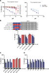
Flunarizine inhibits HCV entry into human hepatocytes in vitro and in vivo. (A) Huh7‐Lunet/hCD81/G‐Luc cells expressing G‐Luc were transfected with F‐Luc‐Jc1. After 4 hours, medium with two‐fold dilutions of flunarizine was added. Measurements of G‐Luc (cell viability) and F‐Luc (RNA replication) were taken after 48 hours. Viruses produced at this time point were used to inoculate target cells where F‐Luc activity was determined 48 hours later (whole life cycle). (B) Huh7‐Lunet/hCD81/G‐Luc cells were inoculated with F‐Luc‐Jc1 and flunarizine or solvent for 4 hours. Infection was measured 48 hours afterwards. (C) Primary human hepatocytes were incubated with the indicated compounds and Jc1 for 6 hours. The supernatant of inoculated cells was collected at 24 hours and 48 hours, and virus infectivity was determined by median tissue culture infective dose. (D) HCV entry reporter mice expressing firefly luciferase in a Cre‐dependent manner were pretreated with the indicated compounds. Subsequently, they were challenged with a Jc1 variant expressing Cre recombinase. HCV‐Cre‐dependent luciferase expression, which reflects HCV cell entry efficiency, was determined 24 hours later. Abbreviation: TCID50, median tissue culture infective dose.
Figure 2
Figure 2
The antiviral activity of flunarizine is HCV strain–dependent, and viral determinants governing susceptibility reside within the E1 and E2 genes. (A) Chimeric Renilla luciferase reporter viruses (left panel) or F‐Luc‐Jc1 or F‐Luc‐JFH‐1 (right panel) were inoculated with two‐fold dilutions of flunarizine into Huh7‐Lunet/hCD81/G‐Luc cells for 4 hours. Luciferase levels were measured 48 hours postinfection. (B) Chimeras between flunarizine‐susceptible Jc1 (GT2a) and flunarizine‐resistant Con1/C3 (GT1b) were created as indicated and tested for their susceptibility to flunarizine using a focus formation unit assay. (C) HCV trans‐complemented particles harboring GT2a or GT2b E1 and E2 glycoproteins of given primary, patient‐derived viruses13 were used to inoculate cells in the presence (two‐fold dilutions) or absence of flunarizine for 4 hours. Infection efficiency was determined 48 hours postinoculation using Renilla luciferase assays. (D) HCV trans‐complemented particles harboring E1 and E2 glycoproteins of representatives of indicated GT2 subtypes2 were used to inoculate cells in the presence of (two‐fold dilutions) or absence of flunarizine. Infection efficiency was determined 48 hours postinoculation by focus formation unit assay.
Figure 3
Figure 3
Two mutations of conserved residues in E1 and one mutation in E2 confer resistance to flunarizine. (A) Jc1 was passaged in Huh7‐Lunet/hCD81/G‐Luc cells in the presence of given doses of flunarizine or DMSO. The resulting virus populations were used to inoculate naive Huh7‐Lunet/hCD81/G‐Luc cells for 4 hours in the presence of 5.25 µM or 10.5 µM of flunarizine or DMSO. The infection was assessed 48 hours later by immune staining of the NS5A protein (green). DNA in the nucleus was counterstained with 4′,6‐diamidino‐2‐phenylindole (blue). (B) Jc1 luciferase reporter virus (WT) or derivatives thereof carrying the indicated point mutations were used to infect Huh7‐Lunet/hCD81/G‐Luc cells in the presence of 0 µM, 0.65 µM, 1.3 µM, 2.6 µM, 5.25 µM, or 10.5 µM of flunarizine for 4 hours. Renilla luciferase activity was measured 48 hours later. (C) Prevalence of indicated amino acids at positions 267 or 289 in HCV E1 across all GT1‐GT7 strains deposited in the HCV database. The total number of analyzed sequences for each genotype is stated in parentheses. (D) Parental or flunarizine‐resistant Jc1 was incubated for 1 hour at 37°C with serial dilutions of given monoclonal antibodies targeting E2 (AP33, CBH23, or HC11) or a discontinuous epitope of the E1 and E2 complex (AR4A) before inoculation of Huh7‐Lunet/hCD81/G‐Luc cells for 4 hours at 37oC. After 48 hours, cells were lysed and Renilla reporter activity was measured. Abbreviation: WT, wild type.
Figure 4
Figure 4
Flunarizine inhibits HCV membrane fusion at the plasma membrane. (A) Schematic representation of the experimental procedure. Huh7‐Lunet/hCD81/G‐Luc cells were incubated with 5 nM concanamycin A 1 hour before virus inoculation and throughout the experiment until 4 hours post–virus inoculation. Additional drugs or DMSO were applied as indicated by black bars according to protocols denominated I, II, and III. F‐Luc Jc1 particles were inoculated for 2 hours at 4°C. Virus membrane fusion at the plasma membrane was triggered by washing cells with a pH 5 buffer (or a pH 7 buffer as control) for 5 minutes 1 hour after inoculated cells were shifted to 37°C. In all treatments, cells were incubated another 48 hours at 37°C before infection efficiency was quantified by luciferase assays. (B) F‐Luc‐Jc1‐dependent luciferase expression in cells inoculated according to protocol I, II, or III and with pH 5 or pH 7 buffer treatment. (C) R‐Luc‐Jc1‐dependent luciferase expression in cells inoculated with parental Jc1 (wild type) or with Jc1 derivatives carrying indicated resistance mutations. Infection was conducted according to protocol II, depicted in (A). Abbreviations: ConA, concanamycin A; RLU, relative light units.
Figure 5
Figure 5
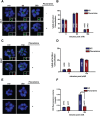
Treatment with flunarizine perturbs DiD‐HCV endosomal membrane fusion. Huh‐7.5 organoids were incubated with 10 μM flunarizine or 10% Dulbecco's modified Eagle's medium alone for 1 hour, infected with DiD‐HCV for 1 hour at 4oC, shifted to 37oC for the indicated times, fixed, and probed for tight junction protein zona occludins‐1 (A) or early endosomal marker EEA1 (C). (A) Tight junction region is shown in the inset. (Left) Zona occludins‐1 (green), (right) DiD‐HCV (red). (B) Quantitation of (A). (C) Arrows indicate DiD‐HCV particles enlarged in insets. (Left) EEA1 (green), (right) DiD‐HCV (red). DiD‐HCV colocalization with EEA1 antibody is indicated by dashed arrow. (D) Quantitation of (C). (E) DiD fluorescence over EEA1 time course. (F) Average DiD fluorescence per cluster in (E). n = total DiD signal (B,D) or total clusters (E); mean ± standard deviation. ***P < 0.001. Abbreviations: VC, vehicle control; ZO‐1, zona occludins‐1.
Figure 6
Figure 6
Flunarizine‐resistant HCV is cross‐resistant to phenothiazines and pimozide, and antiviral activity is not modulated by Ca2+ sequestration. (A) Huh7‐Lunet/hCD81/G‐Luc cells were infected with F‐Luc‐Jc1 in the presence of increasing concentrations of flunarizine either alone or together with the intracellular calcium chelator 1,2‐bis(o‐aminophenoxy)ethane‐N,N,N′,N′‐tetraacetic acid tetra‐(acetoxymethyl)ester or the extracellular calcium chelator ethylene glycol tetraacetic acid. After 2 hours, the viruses and compounds were washed away with phosphate‐buffered saline twice and fresh medium was added. Cells were lysed and the firefly levels measured 48 hours postinfection. (B) Huh7‐Lunet/hCD81/G‐Luc cells were infected with parental Jc1 Renilla reporter viruses (WT) or with the flunarizine‐resistant Jc1 variant (M267V/Q289H/M405T/I757T) for 4 hours in the presence of serially diluted mibefradil, penfluridol, or NiCl2. Medium was changed, and 48 hours later cells were lysed and measured for Renilla luciferase activity. (C) Huh7‐Lunet/hCD81/G‐Luc cells were pretreated for 1 hour with increasing concentrations of methyl‐β‐cyclodextrin to deplete membrane cholesterol. After washing with phosphate‐buffered saline, cells were inoculated with given Renilla reporter viruses for 4 hours. Cells were lyzed and Renilla levels measured 48 hours postinoculation. Abbreviations: BAPTA, 1,2‐bis(o‐aminophenoxy)ethane‐N,N,N′,N′‐tetraacetic acid tetra‐(acetoxymethyl)ester; EGTA, ethylene glycol tetraacetic acid; WT, wild type.
Figure 7
Figure 7
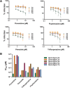
Fluphenazine, pimozide, and trifluoperazine preferentially inhibit HCV infection in a genotype‐dependent fashion but are more active against other genotypes than flunarizine. (A) Huh7‐Lunet/hCD81/G‐Luc cells were inoculated with Renilla luciferase reporter virus chimeras of GT2a (Jc1), GT3a (S52), GT5a (SA13), GT6a (HK6a), or GT7a (QC69) together with two‐fold dilutions of flunarizine, fluphenazine, pimozide, or trifluoperazine. After 48 hours cells were lyzed and measured for Renilla luciferase activity. Means and standard deviations of three independent experiments are given. (B) The calculated IC50 of each compound against each indicated genotype represented in (A) was plotted for comparison.

Comment in

References

    1. Scheel TK, Rice CM. Understanding the hepatitis C virus life cycle paves the way for highly effective therapies. Nat Med 2013;19:837‐849. - PMC - PubMed
    1. Smith DB, Bukh J, Kuiken C, Muerhoff AS, Rice CM, Stapleton JT, et al. Expanded classification of hepatitis C virus into 7 genotypes and 67 subtypes: updated criteria and genotype assignment web resource. Hepatology 2014;59:318‐327. - PMC - PubMed
    1. Lauer GM, Walker BD. Hepatitis C virus infection. N Engl J Med 2001;345:41‐52. - PubMed
    1. Pawlotsky JM. New hepatitis C therapies: the toolbox, strategies, and challenges. Gastroenterology 2014;146:1176‐1192. - PubMed
    1. Mohd Hanafiah K, Groeger J, Flaxman AD, Wiersma ST. Global epidemiology of hepatitis C virus infection: new estimates of age‐specific antibody to HCV seroprevalence. Hepatology 2013;57:1333‐1342. - PubMed

Publication types