. 2015 Nov;66(21):6697-714.
doi: 10.1093/jxb/erv377. Epub 2015 Aug 5.

Xanthomonas campestris cell-cell signalling molecule DSF (diffusible signal factor) elicits innate immunity in plants and is suppressed by the exopolysaccharide xanthan

Affiliations

Xanthomonas campestris cell-cell signalling molecule DSF (diffusible signal factor) elicits innate immunity in plants and is suppressed by the exopolysaccharide xanthan

Akanksha Kakkar et al. J Exp Bot. 2015 Nov.

Abstract

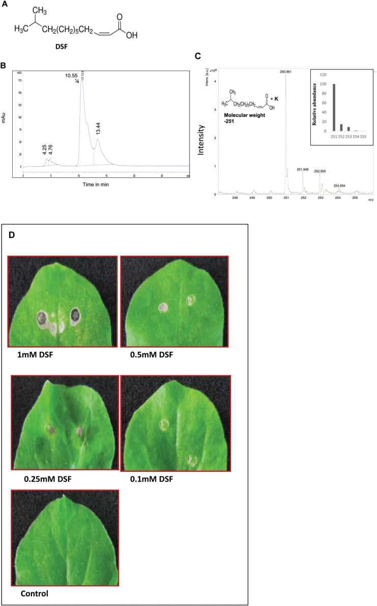
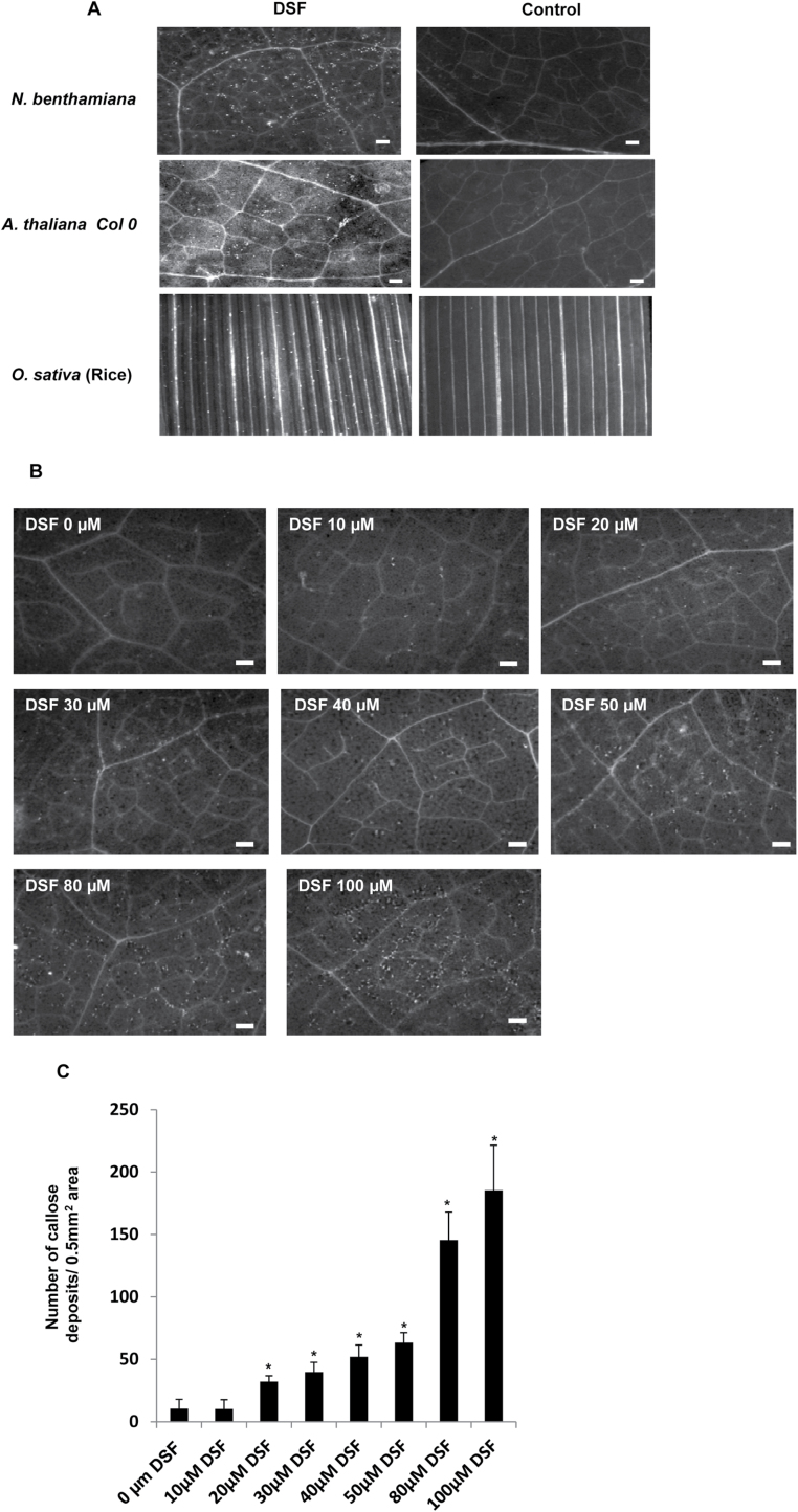
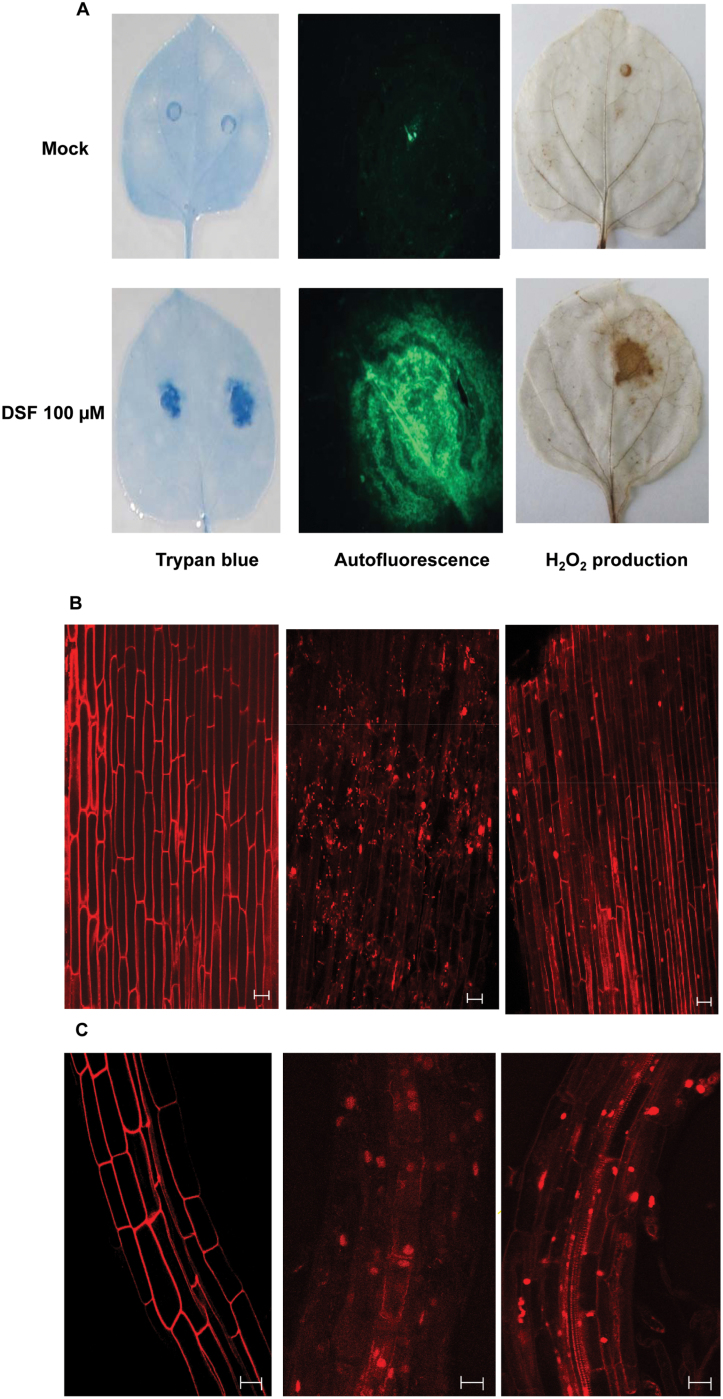
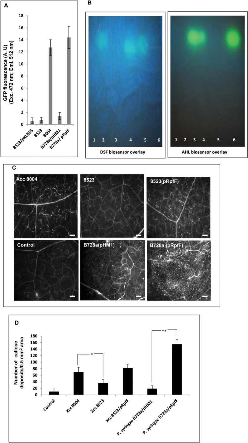
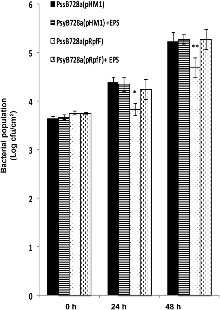
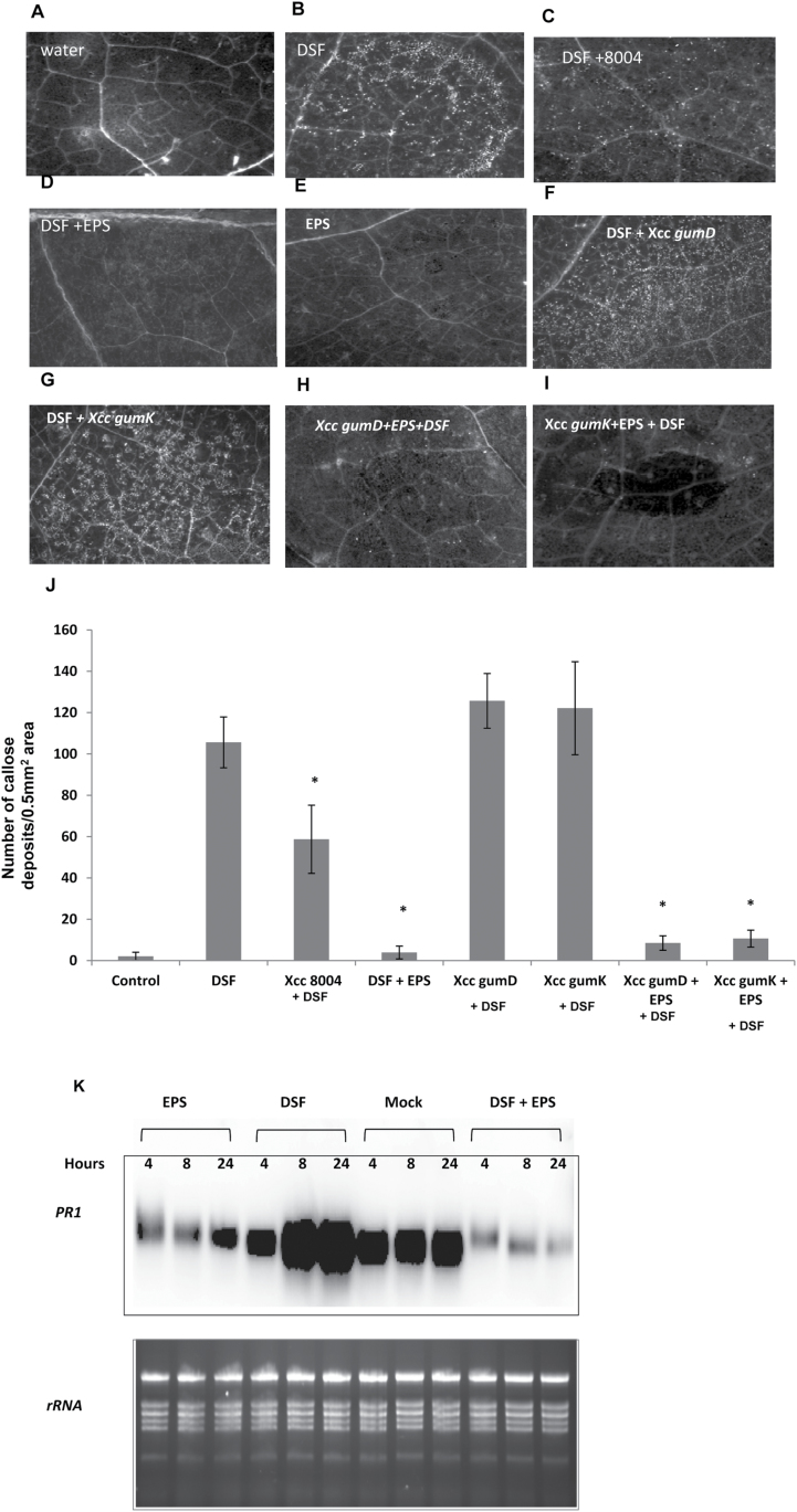
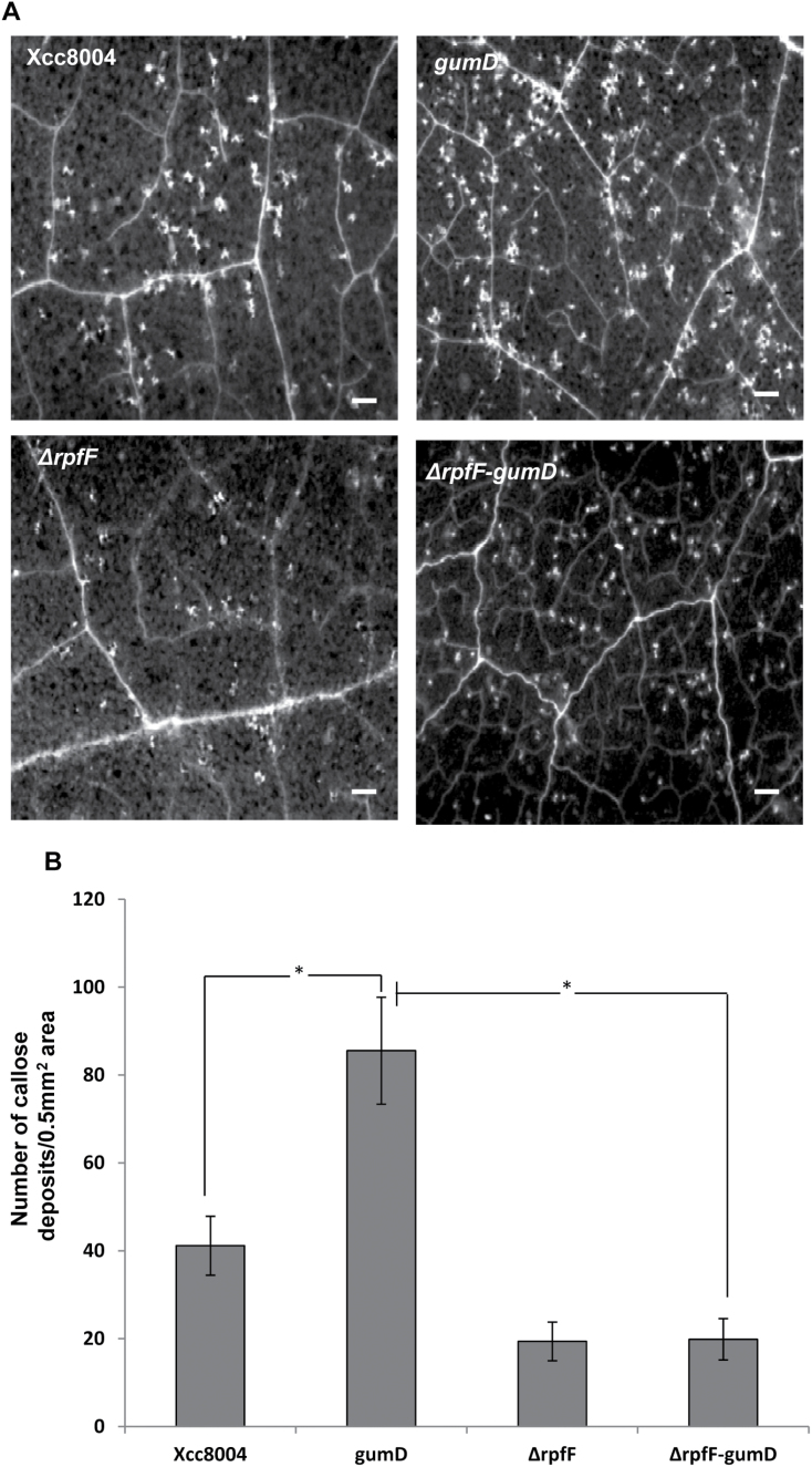
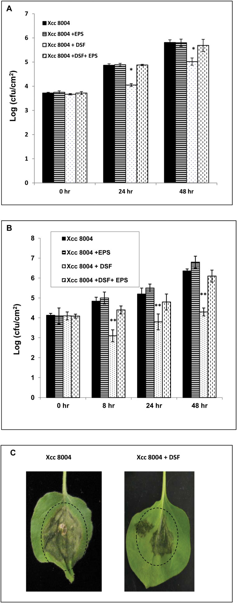
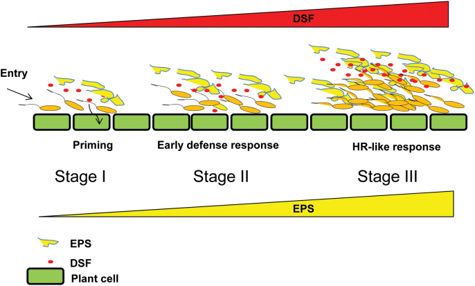
Several secreted and surface-associated conserved microbial molecules are recognized by the host to mount the defence response. One such evolutionarily well-conserved bacterial process is the production of cell-cell signalling molecules which regulate production of multiple virulence functions by a process known as quorum sensing. Here it is shown that a bacterial fatty acid cell-cell signalling molecule, DSF (diffusible signal factor), elicits innate immunity in plants. The DSF family of signalling molecules are highly conserved among many phytopathogenic bacteria belonging to the genus Xanthomonas as well as in opportunistic animal pathogens. Using Arabidopsis, Nicotiana benthamiana, and rice as model systems, it is shown that DSF induces a hypersensitivity reaction (HR)-like response, programmed cell death, the accumulation of autofluorescent compounds, hydrogen peroxide production, and the expression of the PATHOGENESIS-RELATED1 (PR-1) gene. Furthermore, production of the DSF signalling molecule in Pseudomonas syringae, a non-DSF-producing plant pathogen, induces the innate immune response in the N. benthamiana host plant and also affects pathogen growth. By pre- and co-inoculation of DSF, it was demonstrated that the DSF-induced plant defence reduces disease severity and pathogen growth in the host plant. In this study, it was further demonstrated that wild-type Xanthomonas campestris suppresses the DSF-induced innate immunity by secreting xanthan, the main component of extracellular polysaccharide. The results indicate that plants have evolved to recognize a widely conserved bacterial communication system and may have played a role in the co-evolution of host recognition of the pathogen and the communication machinery.

Keywords: Defence suppressor; Nicotiana benthamiana; Xanthomonas campestris pv. campestris; Xanthomonas oryzae pv. oryzae.; elicitor; extracellular polysaccharide; innate immunity; quorum sensing; rice; virulence.


Figures

Fig. 1.
Fig. 1.
Infiltration of synthetic DSF in N. benthamiana induces HR-like symptoms. (A) Chemical structure of DSF, cis-​11-​methyl-​2-​dodecenoic acid. (B and C) Analysis of synthetic DSF by (B) HPLC and (C) MALDI–MS. (B) HPLC separation was achieved with an Agilent C18 (4.6 mm×250 mm×5 μm) column. DSF was eluted with water in methanol (20:80, v/v, with 0.1% formic acid) at a flow rate of 0.8ml min–1 and was detected at 220nm (retention time 10.55) as described in the Materials and methods. (C) Analysis of the MALDI-MS spectrum was done based on quasimolecular ions. Synthetic DSF gave one quasimolecular ion at 251 m/z (inset), which agrees with the calculated mass of a [M+K] + ion. No fragment ions were present at the applied laser energy. (D) Four-week-old N. benthamiana leaves were infiltrated with different concentrations of synthetic DSF. Control: 1% methanol in water. Browning of the infiltrated region and HR-like symptoms were observed 24h post-infiltration.
Fig. 2.
Fig. 2.
DSF induces callose deposition. (A) Infiltration of 100 μM synthetic DSF (cis-11-methyl-2-dodecenoic acid) induces callose deposition in N. benthamiana, Arabidopsis thaliana (Col-0), and rice (Oryzae sativa). Callose deposition was visualized 18h post-infiltration by staining the leaves with aniline blue and examination using an epifluorescence microscope. White dots in these pictures are indicative of callose deposition. (B) DSF induced callose deposition in N. benthamiana leaves in a dose-dependent manner. Nicotiana benthamiana leaves were infiltrated with (left to right) 10, 20, 30, 40, 50, 80, and 100 μM DSF, and control (0 μM; 1% methanol in water), and visualized for callose deposition 18h post-infiltration. Scale bars=500 μm. (C) Average number of callose deposits per 0.5mm2. Error bars represent SD values from four leaves of each plant in three independent experiments. Six microscopic fields from each leaf were analysed. *P<0.01, significant differences between the responses to the DSF treatment compared with the control (indicated by 0 μM) as assessed by Student’s t-test.
Fig. 3.
Fig. 3.
DSF induced cell death in N. benthamiana, rice, and Arabidopsis. (A) DSF induced cell death in N. benthamiana. Leaves of 4-week old plants were infiltrated with 100 μM DSF. The leaves were detached at 24h after DSF infiltration and subjected to observation by staining the leaves with trypan blue, an indicator of cell death (left); autofluorescence (centre); and H2O2 production (right). H2O2 accumulation was visualized by staining with diaminobenzidine (DAB), a histochemical reagent for in situ detection of H2O2. Six or more leaves were examined for each condition, and representative fields are shown. (B and C) DSF induced cell death in rice and Arabidopsis roots. Isolated roots were treated with DSF, cellulase, and control (buffer-treated) for 16h, stained with propidium iodide (PI), and examined under a confocal microscope. Cellulase (a potential DAMP) from Aspergilus niger was used as a positive control. (B) From left to right, rice root tips, 1–2cm long, from 3- to 4-day-old seedlings were treated with control (methanol in water), 100 μM DSF, and cellulase (0.2mg ml–1) from A. niger. (C) From left to right, Arabidopsis roots treated with water control, DSF (100 μM), and cellulase. Treatment with water control did not induce cell death (intake of PI), whereas treatment with either DSF or cellulase induced cell death. Similar results were obtained in at least three independent experiments. Scale bars=20 μm.
Fig. 4.
Fig. 4.
Production of DSF is associated with induced callose deposition. (A) Assay for DSF production by different bacterial strains using the Xcc DSF biosensor 8523 (pKLN55) in which the DSF-responsive promoter from the endoglucanase gene is cloned upstream of an eGFP reporter (Peng:GFP). DSF extracted from the cell-free culture supernatant of Xcc8004 (wild type, DSF+); 8523 (DSF); Pseudomonas syringae pv. syringae PssB728a (pHM1) (wild-type strain harbouring the empty vector); and PssB728a (pRpfF) (Pss harbouring the Xcc DSF synthetic gene rpfF). An increase in GFP fluorescence (represented on the y-axis) compared with the control (extracts from the Xcc8523; rpfF mutant) indicates the amount of DSF produced by different strains. (B) TLC analysis for the production of DSF and AHL in Xcc and PssB728a. TLC was performed with crude ethyl acetate extracts isolated from the culture supernatant of different strains of Xcc and Pss. DSF (left panel) and AHL (right panel) were detected by an overlay of the Xcc DSF indicator strain 8523 (pKLN55) and the E. coli AHL biosensor (JB524) on TLC plates. The photograph was taken with the UV light source oriented from the top of the TLC plates. Column 1, Xcc8523 (DSF-deficient mutant); column 2, Xcc8004 (Xcc wild-type strain); column 3, PssB728a (pHM1); column 4, PssB728a (pRpfF); column 5, synthetic DSF (20 μM cis-11-methyl-2-dodecenoic acid); column 6, synthetic AHL (10 μM N-3-oxo-hexanoyl-dl-homoserine lactone; 3OC6-HSL). Similar results were obtained in three independent experiments. (C) From left to right, callose deposition in N. bethamiana leaves infiltrated with a 1×10⁶ cfu ml–1 suspension of different bacterial strains: wild-type Xcc8004, 8523, 8523 (pRpfF), water control, PssB728a (pHM1), and B728a (pRpfF). Callose deposition was visualized by staining with aniline blue and examined using a stereo fluorescence microscope 24h post-inoculation. White dots in these pictures are indicative of callose deposition. (D) Average number of callose deposits per 0.5mm2 area. Error bars represent SD values from three leaves of each plant from three independent experiments. Six microscopic fields from each leaf were analysed. Differences between the responses to Xcc8523 (DSF mutant; indicated by *P<0.01) and the P. syringae B728a wild-type strain harbouring the Xcc DSF synthase rpfF (indicated by **P<0.001) compared with the wild-type strains of Xcc8004 and P. syringae B728a were significant as assessed by Student’s t-test.
Fig. 5.
Fig. 5.
Production of DSF in Pseudomonas syringae pv. syringae (Pss) reduces growth in N. benthamiana leaves. Leaves of 4-week-old N. benthamiana were infiltrated with wild-type Pss harbouring the plasmid containing the DSF synthase (pRpfF) or the empty vector (pHM1) alone or co-inoculated with Xcc EPS (xanthan; 0.5mg ml–1). The bacterial population was measured at 0, 24, and 48h post-inoculation from six 1cm2 leaf disc areas around the infiltration zone. Values presented are the average log (cfu cm–1) from six leaves (two independent experiments). A significantly different population of bacteria compared with the wild-type Pss harbouring the empty vector control (pHM1) based on a pairwise Student’s t-test is indicated with either one or two asterisks: *P≤0.05; **P≤0.02.
Fig. 6.
Fig. 6.
DSF-induced callose deposition was suppressed by either the wild-type Xcc8004 or the EPS xanthan. (A–I) Callose deposition in N. benthamiana leaves after inoculation with water control (A), 100 μM DSF (B), and EPS (E; xanthan; 0.5mg ml–1) alone or co-inoculation of DSF with either Xcc8004 (C), EPS (D; xanthan), Xcc gumD (F), Xcc gumK (G), Xcc gumD+EPS (H), or Xcc gumK+EPS (I). Callose deposition was visualized by staining with aniline blue and examined using a stereo fluorescence microscope 24h post-inoculation. For co-inoculation experiments with Xcc strains, a bacterial suspension of 1×10⁶ cfu ml–1 was used. (J) Average number of callose deposits per 0.5mm2 area of N. benthamiana leaves inoculated with control (water) and DSF alone, or co-inoculation of DSF with either Xcc8004, EPS, Xcc gumD amd gumK (Xcc mutants deficient in EPS production), or Xcc gumD and gumK mutants supplemented with EPS (gumD and gumK+ EPS). Error bars represent SD values from three leaves of each plant and three independent experiments. Six microscopic fields from each leaf were analysed. * indicates (P<0.001) significantly lower callose deposits compared with leaves treated with DSF alone as determined by two-tailed Student’s t-test. (K) RNA gel blot analysis of PR-1 gene expression in N. benthamiana leaves infiltrated with water (mock), DSF (100 μM), EPS (xanthan, 0.5mg ml–1) alone or co-infiltration with DSF+EPS. RNA was isolated at the indicated times for RNA gel blot analysis. Data shown are representative of those obtained from three independent experiments.
Fig. 7.
Fig. 7.
Callose deposition induced by different strains of Xcc. (A) N. benthamiana leaves were infiltrated with a 1×10⁶ cfu ml–1 suspension of different Xcc strains; Xcc8004 (wild type), gumD (xanthan-deficient mutant), ΔrpfF (DSF-deficient rpfF deletion mutant), and the ΔrpfF-gumD double mutant. Callose deposition was visualized by staining with aniline blue and examined using a stereo fluorescence microscope 24h post-inoculation. White dots in these pictures are indicative of callose deposition. Scale bars=500 μm. (B) Average number of callose deposits per 0.5mm2 area. Error bars represent SD values from three leaves of each plant and three independent experiments. Six microscopic fields from each leaf were analysed. * indicates (P<0.001) significantly different callose deposits induced by the Xcc gumD mutant compared with either the wild-type Xcc8004 strain or the ΔrpfF-gumD double mutant as determined by two-tailed Student’s t-test.
Fig. 8.
Fig. 8.
Pre-treatment or co-infiltration with DSF inhibits wild-type Xcc8004 growth in N. benthamiana leaves. (A) Leaves of 4-week-old plants were pre-infiltrated with either control buffer (black bars), EPS (xanthan; 0.5mg ml–1), DSF (100 μM), or DSF+EPS (xanthan; 0.5mg ml–1) by syringe infiltration 16h prior to inoculation with a 107 cfu ml–1 suspension of the wild-type Xcc8004 strain. Bacterial populations were measured at 0, 24, and 48h post-inoculation. Values presented are average log (cfu cm–2) from six leaves (three leaves each from two independent experiments). * indicates (P≤0.05) a significantly lower bacterial population in the DSF-pre-inoculated leaves compared with the leaves inoculated with the wild-type Xcc8004 strain alone based on a pairwise Student’s t-test. (B) Bacterial growth assay in N. benthamiana leaves co-infiltrated with the Xcc8004 wild-type strain with either water control (black bars), DSF (100 μM), EPS (xanthan; 0.5mg ml–1), or DSF+EPS (xanthan). The bacterial population was measured at 0, 8, 24, and 48h after inoculation. Values presented are average log (cfu ml–1) from six leaves (three leaves each from two independent experiments). * indicates (P≤0.001) a significantly lower bacterial population in the DSF-co-inoculated leaves compared with the leaves inoculated with wild-type Xcc alone based on a pairwise Student’s t-test. (C) Photographs of representative leaves from the co-inoculation experiments 4 d post-inoculation. N. benthamiana leaves exhibit water soaking-like disease symptoms when inoculated with the Xcc8004 wild-type strain (shown in a dotted circle). Leaves co-inoculated with the Xcc8004 wild-type strain+DSF exhibited less vigorous water soaking symptoms compared with leaves treated with the Xcc8004 strain alone.
Fig. 9.
Fig. 9.
Proposed model for functional interplay between diffusible signalling factor (DSF) and extracellular polysaccharide (EPS) in Xanthomonas–plant interaction. At the initial stage of infection and colonization (stage I), Xcc gains entry through hydathodes or stomata, and colonizes in the xylem vessel. At this stage (at low cell density; stage I), the production of DSF and EPS is low. At lower concentrations of DSF (presumably ≤10 μM), DSF may be involved in priming (sensitization) plants for MTI (MAMP-triggered immunity) mediated by MAMPs such as flagellin or LPS (lipopolysaccharide). MTI is further suppressed by Type III secretion system effectors. At stage II, there is increase in Xcc cell number, which is associated with increased production of DSF and EPS. An increased DSF level (≥20 μM) induces an early plant defence response (callose deposition), which is suppressed by EPS. At a late stage of infection (stage III), there is a further increase in cell density due to growth of Xcc in planta. Due to high cell density, a high level of DSF is produced (50–100 μM). This may lead to a further increase in the production of EPS, a virulence-associated factor positively regulated by DSF. A high EPS level can suppress the plant defence response provoked by DSF including early HR-like symptoms.

References

    1. Adam L, Somerville SC. 1996. Genetic characterization of five powdery mildew disease resistance loci in Arabidopsis thaliana . The Plant Journal 9, 341–356. - PubMed
    1. Altschul SF, Madden TL, Schaffer AA, Zhang J, Zhang Z, Miller W, Lipman DJ. 1997. Gapped BLAST and PSI-BLAST: a new generation of protein database search programs. Nucleic Acids Research 25, 3389–3402. - PMC - PubMed
    1. Aparna G, Chatterjee A, Sonti RV, Sankaranarayanan R. 2009. A cell wall-degrading esterase of Xanthomonas oryzae requires a unique substrate recognition module for pathogenesis on rice. The Plant Cell 21, 1860–1873. - PMC - PubMed
    1. Aslam SN, Newman MA, Erbs G, et al. 2008. Bacterial polysaccharides suppress induced innate immunity by calcium chelation. Current Biology 18, 1078–1083. - PubMed
    1. Barber CE, Tang JL, Feng JX, Pan MQ, Wilson TJG, Slater H, Dow JM, Williams P, Daniels MJ. 1997. A novel regulatory system required for pathogenicity of Xanthomonas campestris is mediated by a small diffusible signal molecule. Molecular Microbiology 24, 555–566. - PubMed

Publication types

MeSH terms