Clinical Trial
. 2015 Oct;45(10):2858-72.
doi: 10.1002/eji.201445404. Epub 2015 Aug 28.

Progesterone promotes maternal-fetal tolerance by reducing human maternal T-cell polyfunctionality and inducing a specific cytokine profile

Affiliations
Clinical Trial

Progesterone promotes maternal-fetal tolerance by reducing human maternal T-cell polyfunctionality and inducing a specific cytokine profile

David Lissauer et al. Eur J Immunol. 2015 Oct.

Abstract

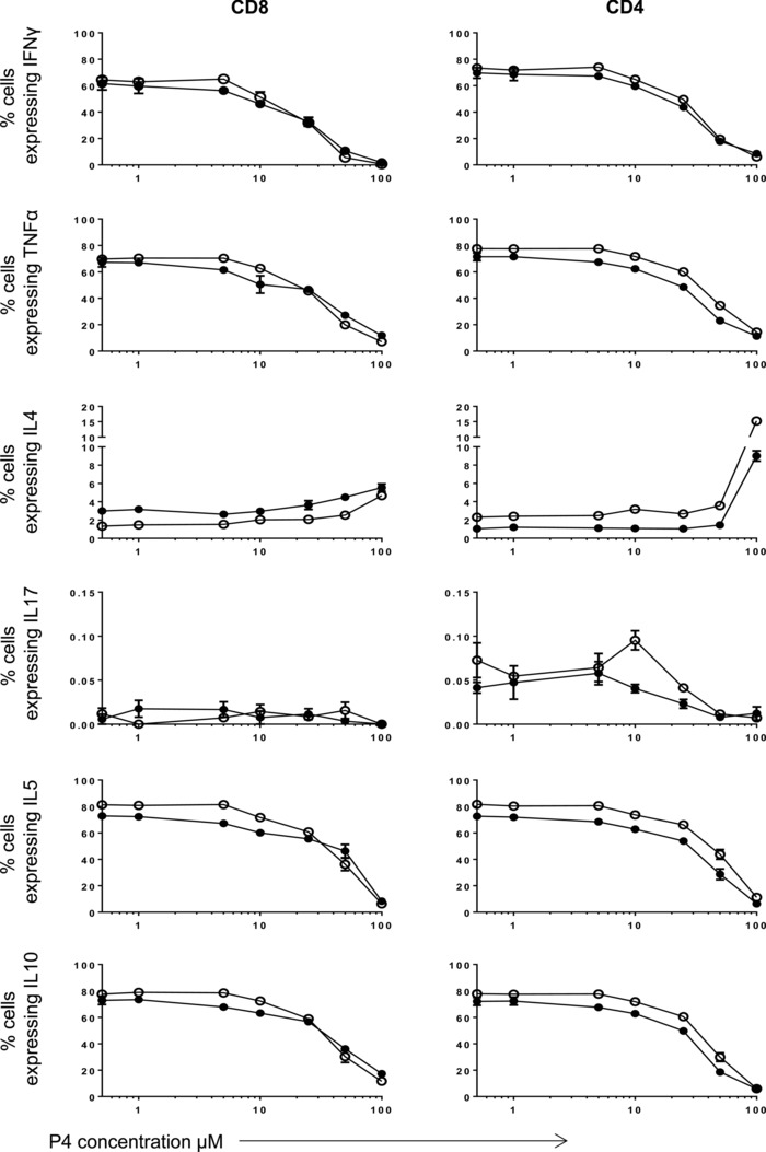
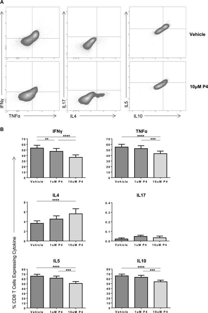
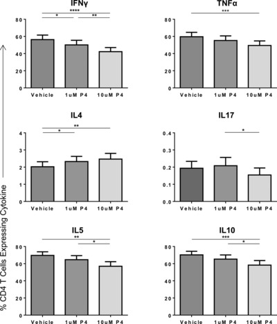
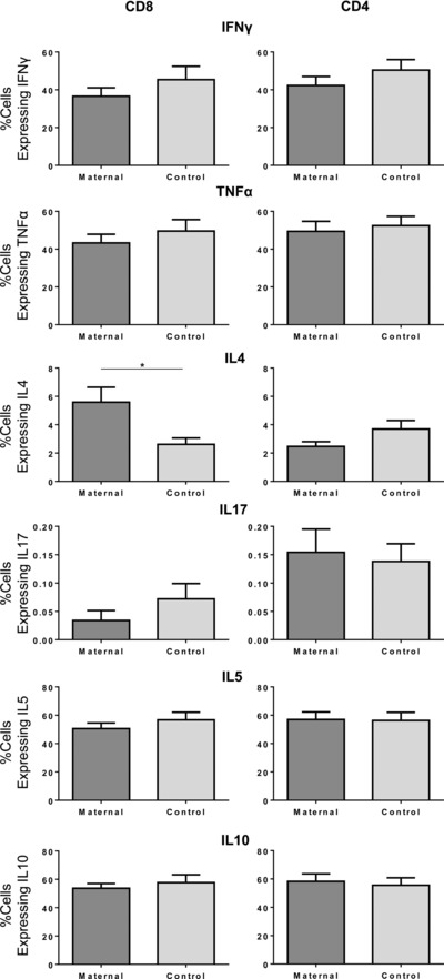
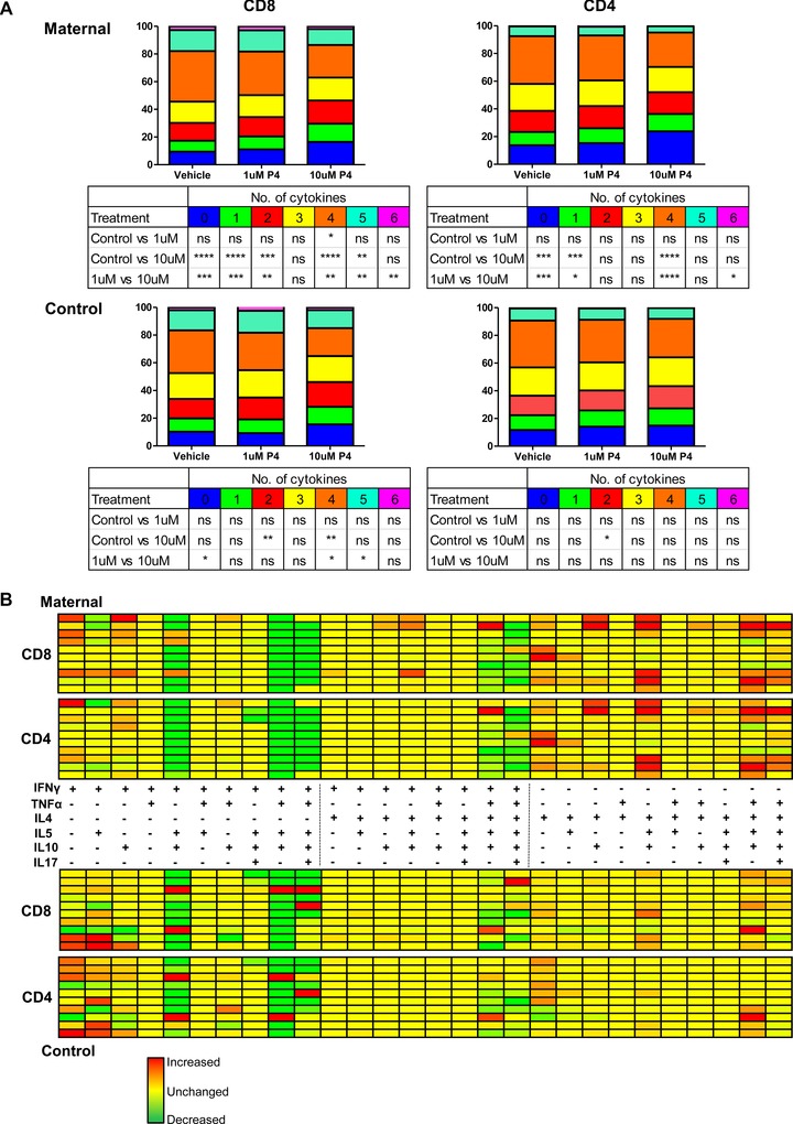
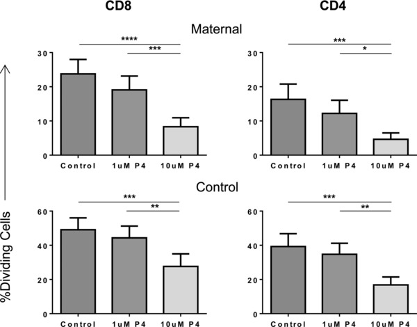
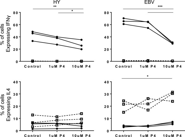
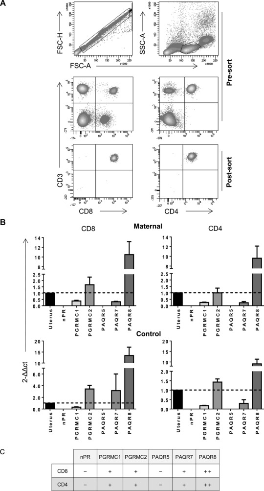
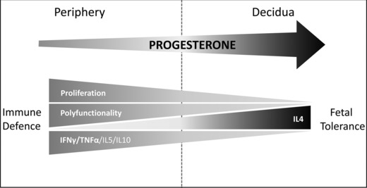
Progesterone is a steroid hormone essential for the maintenance of human pregnancy, and its actions are thought to include promoting maternal immune tolerance of the semiallogenic fetus. We report that exposure of maternal T cells to progesterone at physiological doses induced a unique skewing of the cytokine production profile of CD4(+) and CD8(+) T cells, with reductions not only in potentially deleterious IFN-γ and TNF-α production but also in IL-10 and IL-5. Conversely, production of IL-4 was increased. Maternal T cells also became less polyfunctional, focussing cytokine production toward profiles including IL-4. This was accompanied by reduced T-cell proliferation. Using fetal and viral antigen-specific CD8(+) T-cell clones, we confirmed that this as a direct, nonantigen-specific effect. Yet human T cells lacked conventional nuclear progesterone receptors, implicating a membrane progesterone receptor. CD4(+) and CD8(+) T cells responded to progesterone in a dose-dependent manner, with subtle effects at concentrations comparable to those in maternal blood, but profound effects at concentrations similar to those at the maternal-fetal interface. This characterization of how progesterone modulates T-cell function is important in understanding the normal biology of pregnancy and informing the rational use of progesterone therapy in pregnancies at risk of fetal loss.

Keywords: Human; IL-4; Maternal-fetal tolerance; Progesterone; T cell.


Figures

Figure 1
Figure 1
The effect of progesterone on cytokine production by human maternal CD8+ and CD4+ T cells is dose‐dependent. PBMCs from healthy maternal (●) and control (○) donors (n = 1, experiment in triplicate) were treated with PHA and increasing concentrations of progesterone (P4) from 0.5 to 100 μM. The effect of progesterone treatment on the production of IFN‐γ, TNF‐α, IL‐4, IL‐17, IL‐5, and IL‐10 was measured by flow cytometry. Data are shown as mean + SEM from a single experiment.
Figure 2
Figure 2
Treatment of maternal PBMCs with physiological concentrations of progesterone alters the cytokine expression of CD8+ T cells. PBMCs from healthy maternal donors were treated with PHA and either DMSO (vehicle), or 1 or 10 μM progesterone. The effect of progesterone treatment on production of IFN‐γ, TNF‐α, IL‐4, IL‐17, IL‐5, and IL‐10 was measured by flow cytometry. (A) A representative flow plot of PBMCs from one patient treated with DMSO and 10 μM progesterone (P4) is shown. (B) The cytokine expression of maternal CD8+ T cells overall when treated with different progesterone concentrations or vehicles is shown as mean + SEM of 13 donors. *p ≤ 0.05, **p ≤ 0.01, ***p ≤ 0.001, ****p ≤ 0.0001, one‐way ANOVA, repeated measures, and Bonferroni multiple comparison.
Figure 3
Figure 3
Treatment of maternal PBMCs with physiological concentrations of progesterone alters the cytokine profile of CD4+ T cells. PBMCs from maternal donors were treated with PHA and either DMSO (vehicle), or 1 or 10 μM progesterone and the effect of progesterone treatment on production of IFN‐γ, TNF‐α, IL‐4, IL‐17, IL‐5, and IL‐10 was measured by flow cytometry. The cytokine profile of maternal CD4+ T cells overall when treated with different progesterone concentrations or vehicle is shown as mean + SEM of 13 donors. *p ≤ 0.05, **p ≤ 0.01, ***p ≤ 0.001, ****p ≤ 0.0001, one‐way ANOVA, repeated measures, and Bonferroni multiple comparison test.
Figure 4
Figure 4
The percentage of CD8+ T cells expressing IL‐4 is significantly higher in maternal subjects compared with that of controls following treatment with 10 μM progesterone. The cytokine profile of maternal CD4+ and CD8+ T cells (dark gray, n = 13) was compared with that of control cells (light gray, n = 11) where PBMCs were treated with PHA and 10 μM progesterone, as measured by flow cytometry. Data shown as mean + SEM of indicated numbers of donors. *p ≤ 0.05, unpaired t‐test.
Figure 5
Figure 5
Progesterone reduces the polyfunctional cytokine profile of CD8+ and CD4+ maternal T cells and favors a more IL‐4 dominant profile. (A) A Boolean gating strategy was established to look at the percentage of CD8+ and CD4+ T cells expressing from zero to six cytokines from both the maternal (top) and control (bottom) donors (n = 10 of each group), where PBMCs had been treated with PHA and either DMSO (vehicle), or 1 or 10 μM progesterone. *p ≤ 0.05, **p ≤ 0.01, ***p ≤ 0.001, ****p ≤ 0.0001, one‐way ANOVA, repeated measures, and Bonferroni multiple comparison test. (B) The same strategy was then used to look at the percentage of CD8+ and CD4+ T cells expressing each individual cytokine and every possible combination up to six cytokines. The differential absolute expression between the vehicle and 10 μM progesterone was used to create a heat map indicating the change in cytokine combinations when maternal or male PBMCs were treated with PHA and 10 μM progesterone. The change in cytokine expression following progesterone treatment is represented by a range of colors of varying green (increase) and red (decrease) intensity, denoting the percentage change in cytokine production with progesterone treatment. The map has been laid out in three sections; the left section is combination of cytokines including IFN‐γ but excluding IL‐4, the middle section is combination including both IFN‐γ and IL‐4, and the right section is inclusive of IL‐4 but excludes IFN‐γ.
Figure 6
Figure 6
Progesterone treatment reduces the proliferation of CD8+ and CD4+ T cells. PBMCs from maternal and control donors were labeled with CellTrace™, treated with PHA and either vehicle, or 1 or 10 μM progesterone. The percentages of dividing maternal and control CD8+ and CD4+ T cells overall when treated with different progesterone concentrations or vehicle were then measured by flow cytometry and shown as mean + SEM of five donors in each group. *p ≤ 0.05, **p ≤ 0.01, ***p ≤ 0.001, ****p ≤ 0.0001, one‐way ANOVA, repeated measures, and Bonferroni multiple comparison test.
Figure 7
Figure 7
Treatment of antigen‐specific CD8+ T‐cell clones with progesterone suggests that changes in the cytokine profile of these cells occur through both direct and indirect effects. Overall effects of progesterone treatment on the percentage of HY antigen‐specific CD8+ T cells expressing IFN‐γ (top) and IL‐4 (bottom) following incubation with target cells loaded with HY‐specific peptide (●) or irrelevant peptide (□) (left) and on EBV clones specific for the GLC peptide incubated with target cells loaded with GLC peptide (●) or knock out cells unable to express GLC peptide (□) (right). Three independent HY clones, and three independent EBV clones were tested over three experiments; *p ≤ 0.05, **p ≤ 0.01, ***p ≤ 0.001, ****p ≤ 0.0001, one‐way ANOVA, repeated measures, and Bonferroni multiple comparison test.
Figure 8
Figure 8
The expression of membrane and nuclear progesterone receptors on CD8+ and CD4+ T cells. (A) PBMCs isolated from maternal and control donors were sorted into pure populations of CD8+ and CD4+ T cells and purity was checked (n = 4). (B) RNA was extracted from the CD8+ and CD4+ T‐cell populations, and real‐time PCR was used to determine the expression of the nuclear progesterone receptors (nPR), progestin and adipoQ receptor family members 5, 7, and 8 (PAQR5, 7, and 8), and the progesterone receptor membrane component members 1 and 2 (PGRMC1 and 2). Human uterus cDNA was used as a positive control and GAPDH as the endogenous control/housekeeper. Data shown as mean + SEM of n = 4 samples in each group. (C) Summary of nuclear and membrane progesterone receptor expression levels found on CD8+ and CD4+ T cells relative to the levels in uterus.
Figure 9
Figure 9
Schematic representation of the influence of progesterone concentration on CD4+ and CD8+ T‐cell function during pregnancy. Exposure of maternal T cells to progesterone at concentrations similar to those found at the maternal–fetal interface (right) induced a unique skewing of the cytokine production profile of CD4+ and CD8+ T cells, with reductions in potentially deleterious IFN‐γ and TNF‐α production but also reductions in IL‐10 and IL‐5. Conversely, production of IL‐4 was increased. Maternal T cells became less polyfunctional, with fewer cytokines produced but tending to include IL4. This was accompanied by reduced T‐cell proliferation. CD4+ and CD8+ T cells responded to progesterone in a dose‐dependent manner, with only subtle effects at concentrations comparable to those in maternal peripheral blood (left) facilitating continued host defense. Thus, the tolerogenic effects are localized to the maternal–fetal interface where higher concentrations of progesterone are found (right).

References

    1. Lissauer, D. M. , Piper, K. P. , Moss, P. A. and Kilby, M. D. , Fetal microchimerism: the cellular and immunological legacy of pregnancy. Expert Rev. Mol. Med. 2009. 11: e33. - PubMed
    1. Tafuri, A. , Alferink, J. , Moller, P. , Hammerling, G. J. and Arnold, B. , T cell awareness of paternal alloantigens during pregnancy. Science 1995. 270: 630–633. - PubMed
    1. Lissauer, D. , Piper, K. , Goodyear, O. , Kilby, M. D. and Moss, P. A. , Fetal‐specific CD8+ cytotoxic T cell responses develop during normal human pregnancy and exhibit broad functional capacity. J. Immunol. 2012. 189: 1072–1080. - PubMed
    1. Piper, K. P. , McLarnon, A. , Arrazi, J. , Horlock, C. , Ainsworth, J. , Kilby, M. D. , Martin, W. L. et al., Functional HY‐specific CD8+ T cells are found in a high proportion of women following pregnancy with a male fetus. Biol. Reprod. 2007. 76: 96–101. - PubMed
    1. Lissauer, D. , Choudhary, M. , Pachnio, A. , Goodyear, O. , Moss, P. A. and Kilby, M. D. , Cytomegalovirus sero positivity dramatically alters the maternal CD8+ T cell repertoire and leads to the accumulation of highly differentiated memory cells during human pregnancy. Hum. Reprod. 2011. 26: 3355–3365. - PubMed

Publication types