Metallic zinc exhibits optimal biocompatibility for bioabsorbable endovascular stents
- PMID: 26249616
- PMCID: PMC4529538
- DOI: 10.1016/j.msec.2015.07.022
Metallic zinc exhibits optimal biocompatibility for bioabsorbable endovascular stents
Abstract
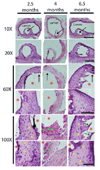
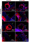
Although corrosion resistant bare metal stents are considered generally effective, their permanent presence in a diseased artery is an increasingly recognized limitation due to the potential for long-term complications. We previously reported that metallic zinc exhibited an ideal biocorrosion rate within murine aortas, thus raising the possibility of zinc as a candidate base material for endovascular stenting applications. This study was undertaken to further assess the arterial biocompatibility of metallic zinc. Metallic zinc wires were punctured and advanced into the rat abdominal aorta lumen for up to 6.5months. This study demonstrated that metallic zinc did not provoke responses that often contribute to restenosis. Low cell densities and neointimal tissue thickness, along with tissue regeneration within the corroding implant, point to optimal biocompatibility of corroding zinc. Furthermore, the lack of progression in neointimal tissue thickness over 6.5months or the presence of smooth muscle cells near the zinc implant suggest that the products of zinc corrosion may suppress the activities of inflammatory and smooth muscle cells.
Keywords: Bioabsorbable; Biocompatible; Corrosion; Hyperplasia; Stent; Zinc.
Copyright © 2015 Elsevier B.V. All rights reserved.
Figures


Similar articles
-
MicroCT and contrast-enhanced microCT to study the in vivo degradation behavior and biocompatibility of candidate metallic intravascular stent materials.Acta Biomater. 2025 Jan 1;191:53-65. doi: 10.1016/j.actbio.2024.11.017. Epub 2024 Nov 17. Acta Biomater. 2025. PMID: 39561850
-
Long-term surveillance of zinc implant in murine artery: Surprisingly steady biocorrosion rate.Acta Biomater. 2017 Aug;58:539-549. doi: 10.1016/j.actbio.2017.05.045. Epub 2017 May 19. Acta Biomater. 2017. PMID: 28532901 Free PMC article.
-
A simplified in vivo approach for evaluating the bioabsorbable behavior of candidate stent materials.J Biomed Mater Res B Appl Biomater. 2012 Jan;100(1):58-67. doi: 10.1002/jbm.b.31922. Epub 2011 Sep 8. J Biomed Mater Res B Appl Biomater. 2012. PMID: 21905215
-
Biodegradable Metals for Cardiovascular Stents: from Clinical Concerns to Recent Zn-Alloys.Adv Healthc Mater. 2016 May;5(10):1121-40. doi: 10.1002/adhm.201501019. Epub 2016 Apr 20. Adv Healthc Mater. 2016. PMID: 27094868 Free PMC article. Review.
-
Coating bioabsorption and chronic bare metal scaffolding versus fully bioabsorbable stent.EuroIntervention. 2009 Dec 15;5 Suppl F:F36-42. doi: 10.4244/EIJV5IFA6. EuroIntervention. 2009. PMID: 22100674 Review.
Cited by
-
Enhanced cytocompatibility and antibacterial property of zinc phosphate coating on biodegradable zinc materials.Acta Biomater. 2019 Oct 15;98:174-185. doi: 10.1016/j.actbio.2019.03.055. Epub 2019 Mar 29. Acta Biomater. 2019. PMID: 30930304 Free PMC article.
-
Blending with transition metals improves bioresorbable zinc as better medical implants.Bioact Mater. 2022 Jun 2;20:243-258. doi: 10.1016/j.bioactmat.2022.05.033. eCollection 2023 Feb. Bioact Mater. 2022. PMID: 35702610 Free PMC article.
-
The biological effects of copper alloying in Zn-based biodegradable arterial implants.Biomater Adv. 2025 Feb;167:214112. doi: 10.1016/j.bioadv.2024.214112. Epub 2024 Nov 8. Biomater Adv. 2025. PMID: 39561579
-
Biodegradable ZnLiCa ternary alloys for critical-sized bone defect regeneration at load-bearing sites: In vitro and in vivo studies.Bioact Mater. 2021 Apr 20;6(11):3999-4013. doi: 10.1016/j.bioactmat.2021.03.045. eCollection 2021 Nov. Bioact Mater. 2021. PMID: 33997489 Free PMC article.
-
Cardiovascular Stents: A Review of Past, Current, and Emerging Devices.Materials (Basel). 2021 May 12;14(10):2498. doi: 10.3390/ma14102498. Materials (Basel). 2021. PMID: 34065986 Free PMC article. Review.
References
-
- Hascoët S, Baruteau A, Jalal Z, Mauri L, Acar P, Elbaz M, Boudjemline Y, Fraisse A. Arch. Cardiovasc. Dis. 2014;107:462–475. - PubMed
-
- Farb A, Heller PF, Shroff S, Cheng L, Kolodgie FD, Carter AJ, Scott DS, Froehlich J, Virmani R. Circulation. 2001;104:473–479. - PubMed
-
- Virmani R, Liistro F, Stankovic G, Di Mario C, Montorfano M, Farb A, Kolodgie FD, Colombo A. Circulation. 2002;106:2649–2651. - PubMed
-
- Carter AJ, Aggarwal M, Kopia GA, Tio F, Tsao PS, Kolata R, Yeung AC, Llanos G, Dooley J, Falotico R. Cardiovasc. Res. 2004;63:617–624. - PubMed
-
- Joner M, Finn AV, Farb A, Mont EK, Kolodgie FD, Ladich E, Kutys R, Skorija K, Gold HK, Virmani R. J. Am. Coll. Cardiol. 2006;48:193–202. - PubMed
Publication types
MeSH terms
Substances
Grants and funding
LinkOut - more resources
Full Text Sources
Other Literature Sources

