

Metachronous tubulovillous and tubular adenomas of the anal canal
- PMID: 26249723
- PMCID: PMC4528720
- DOI: 10.1186/s13000-015-0379-9
Metachronous tubulovillous and tubular adenomas of the anal canal
Abstract
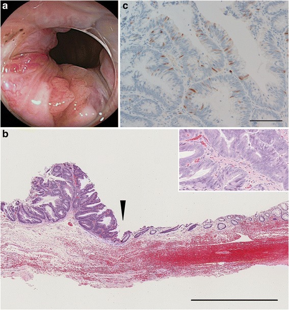
Anal canal adenoma is an extremely rare disease that has the potential to transform into a malignant tumor. We herein presented a rare case of metachronous multiple adenomas of the anal canal. A 48-year-old woman underwent total colonoscopy following a positive fecal blood test. A 9-mm villous polyp arising from the posterior wall of the anal canal was removed by snare polypectomy. Histologically, the tumor was tubulovillous adenoma with high-grade dysplasia and the cut end was negative for tumor cells. Six years later, an elevated lesion, macroscopically five millimeters in size, was detected in the left wall of the anal canal in a follow-up colonoscopy. Local excision of the tumor was performed, and the lesion was pathologically confirmed to be tubular adenoma with high-grade dysplasia limited to the mucosa. The patient is currently alive without any evidence of recurrence for six months after surgery. Although she had a past history of cervical cancer, the multiple tumors arising in the anal canal were unlikely to be related to human papilloma virus infection. Our case report underscores the importance of careful observations throughout colonoscopy to detect precancerous lesions, particularly in anatomically narrow segments.
Figures


Similar articles
-
Tubulovillous adenoma of anal canal: a case report.World J Gastroenterol. 2006 Mar 21;12(11):1780-1. doi: 10.3748/wjg.v12.i11.1780. World J Gastroenterol. 2006. PMID: 16586552 Free PMC article.
-
Tubulovillous adenoma of the anal canal: report of 2 rare cases with review of literature.Ann Diagn Pathol. 2012 Jun;16(3):210-3. doi: 10.1016/j.anndiagpath.2011.01.002. Epub 2011 Mar 29. Ann Diagn Pathol. 2012. PMID: 21447446 Review.
-
Characteristics of metachronous colorectal adenomas found during long-term follow-up: analysis of four subsequent generations of adenoma recurrence.Scand J Gastroenterol. 2009;44(6):736-44. doi: 10.1080/00365520902770078. Scand J Gastroenterol. 2009. PMID: 19277927
-
Predictors of colorectal neoplasia after polypectomy: based on initial and consecutive findings.Neth J Med. 2014 Apr;72(3):139-45. Neth J Med. 2014. PMID: 24846927
-
[Intra-epithelial cancer of the anal canal. Pathogenic study apropos of 5 cases].Gastroenterol Clin Biol. 1990;14(3):224-9. Gastroenterol Clin Biol. 1990. PMID: 2188862 Review. French.
References
Publication types
MeSH terms
LinkOut - more resources
Full Text Sources
Other Literature Sources
Medical
Miscellaneous
